- Giới thiệu
- Giải pháp
- Y sinh
- Các giải pháp sàng lọc trước sinh và sơ sinh
- Xét nghiệm di truyền
- An toàn truyền máu
- Giải pháp xét nghiệm nhanh
- Actim PROM - Test nhanh chẩn đoán rỉ ối
- Actim® Partus – Test nhanh hỗ trợ chẩn đoán nguy cơ sinh non
- Actim Calprotectin – Test nhanh bán định lượng hỗ trợ chẩn đoán viêm ruột
- Actim Pancreatitis: Chẩn đoán Viêm tụy cấp nhanh chóng và chính xác
- Actim CRP - Xét nghiệm nhanh CRP
- Abbott - Xét nghiệm chẩn đoán nhanh bệnh truyền nhiễm
- Xét nghiệm Sinh hóa - Miễn dịch VITROS
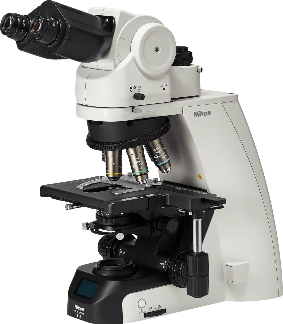
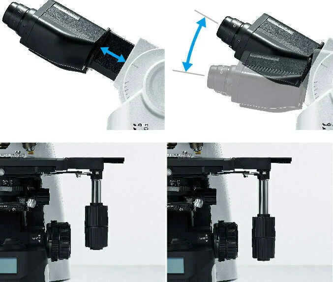
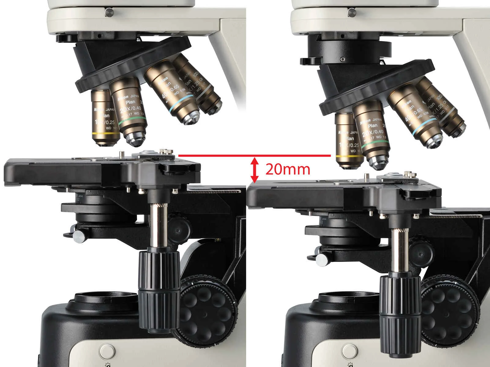
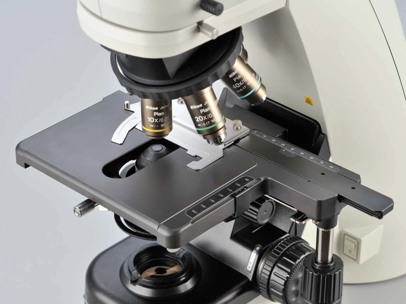
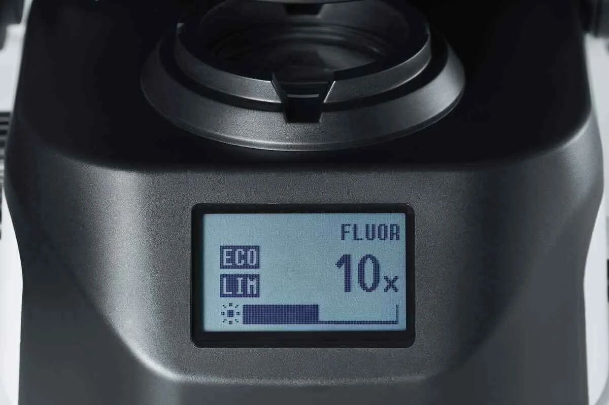
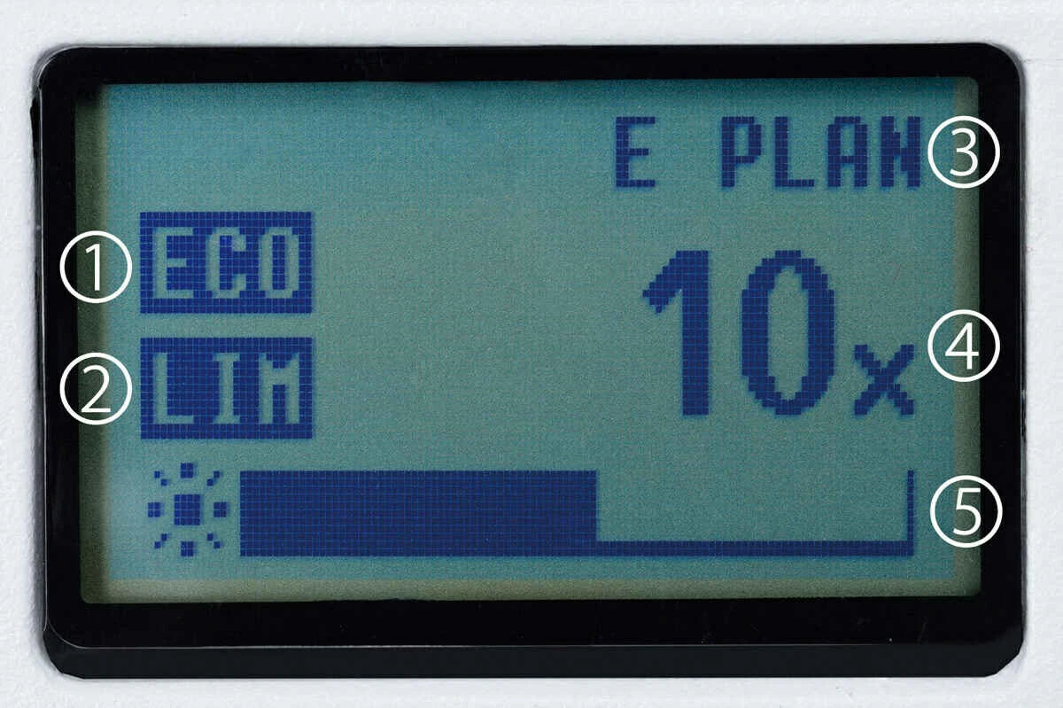
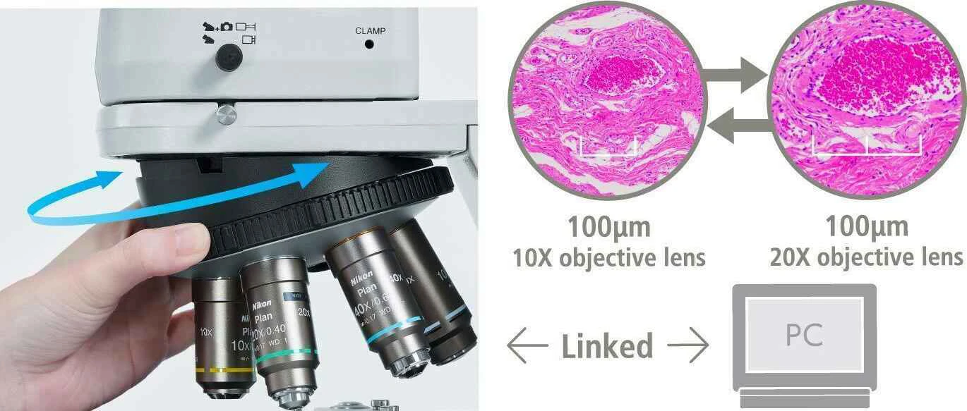
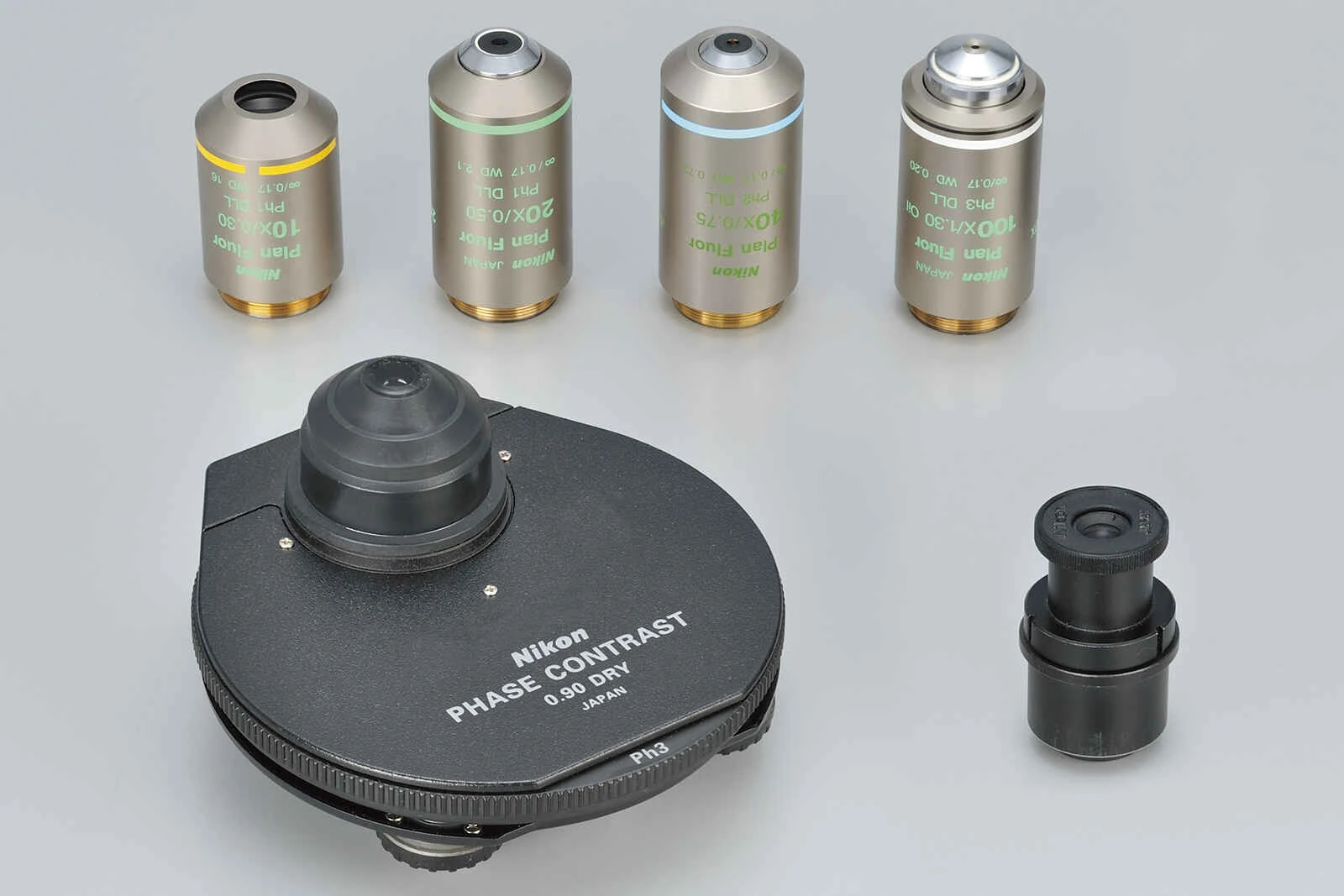
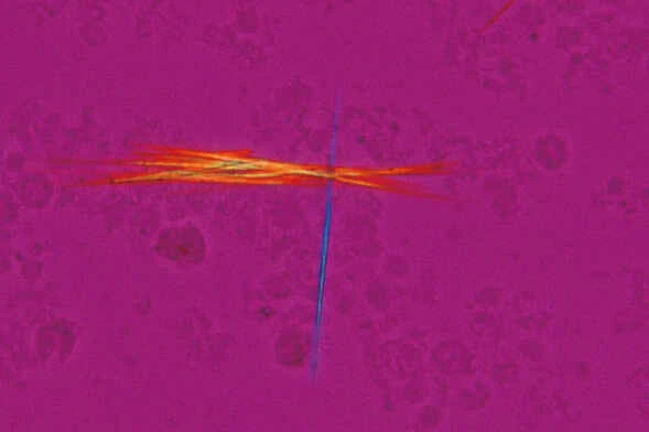
- Kính hiển vi Nikon & Giải pháp hình ảnh
- Sàng lọc ung thư cổ tử cung
- Hóa lý
- Phân tích trong ngành thực phẩm & thức ăn chăn nuôi
- Phân tích cho môi trường
- Nghiên cứu và phát triển ứng dụng cho vi sinh, sinh học, dược phẩm
- Spray Drying & Encapsulation Solutions – Sấy phun và tạo vi nang bảo vệ hoạt chất.
- Freeze Drying Solutions – Đông khô mẫu trong nghiên cứu dược phẩm, công nghệ sinh học
- Melting Point Solutions – Xác định điểm nóng chảy, đánh giá độ tinh khiết của dược phẩm, hóa chất
- Distillation & Evaporation Solutions – Bay hơi quay cô đặc, chiết xuất hợp chất hữu cơ.
- Flash Chromatography Solutions – Phân tách nhanh các hợp chất dược liệu.
- Preparative Chromatography Solutions – Tinh sạch hợp chất bằng sắc ký điều chế.
- Phân tích thành phần hóa và khoáng
- Phân tích thành phần hóa và khoáng trong ngành xi măng
- Phân tích thành phần hóa và khoáng trong ngành khoáng sản
- Phân tích thành phần hóa và khoáng trong ngành ceramic
- Phân tích thành phần hóa và khoáng trong ngành sản xuất sắt thép phủ mạ
- Phân tích thành phần hóa và khoáng trong ngành dầu khí
- Phân tích thành phần hóa và khoáng trong ngành sản xuất Pin
- Phân tích cấu trúc và hình thái vật liệu
- Thiết bị phòng thí nghiệm cơ bản
- Địa không gian
- Y sinh
- Sự kiện
- Tuyển dụng
- Liên hệ
VN
EN