Trusted by pathologists worldwide, Nikon microscopes deliver exceptional image quality for accurate diagnosis in cancer and autoimmune diseases.
From brightfield to digital slide scanning and fluorescence, Nikon provides complete imaging solutions for diagnostics, teaching, and research.
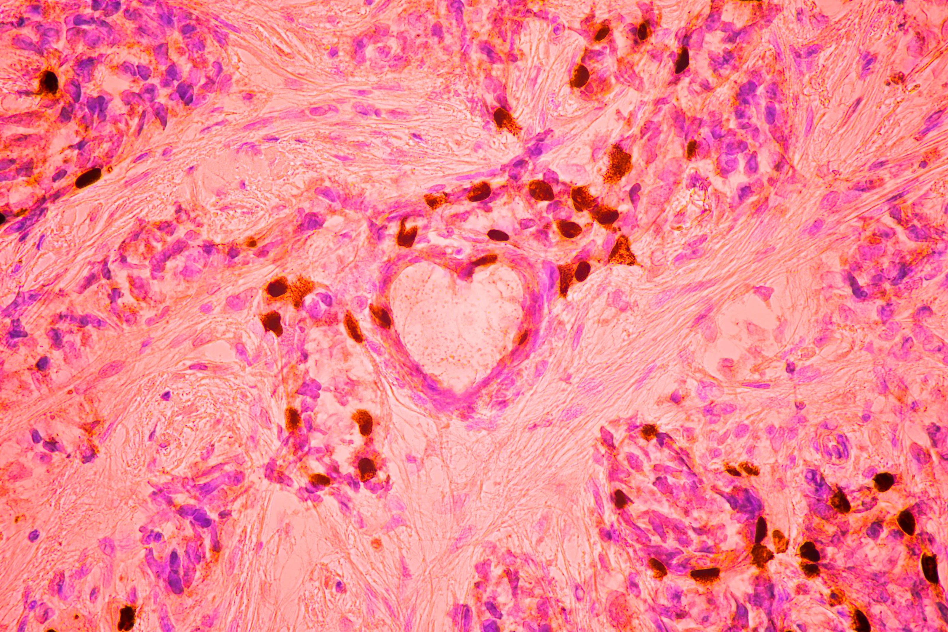
High-resolution image of breast cancer cells captured with a Nikon microscope
Nikon Microscopes – Versatile Solutions for Modern Pathology
In the histopathological diagnostic workflow, Nikon microscopes are used to examine tissue samples stained with Hematoxylin and Eosin (H&E), immunohistochemistry (IHC), PAS, Masson’s Trichrome, Giemsa, Ziehl–Neelsen, or various multicolor fluorescent markers.
H&E and Pap-stained tissue images captured with a Nikon microscope and camera system (Hình 1, Hình 2)
Nikon’s CFI60 optical system and high-performance objective lenses deliver sharp images with high contrast, true-to-life color reproduction, and optimal depth of field — essential features for pathologists to clearly distinguish lesion boundaries, normal vs. abnormal tissue structures, nuclear and cytoplasmic details, degrees of invasion, and the microscopic morphology of cancer cells.
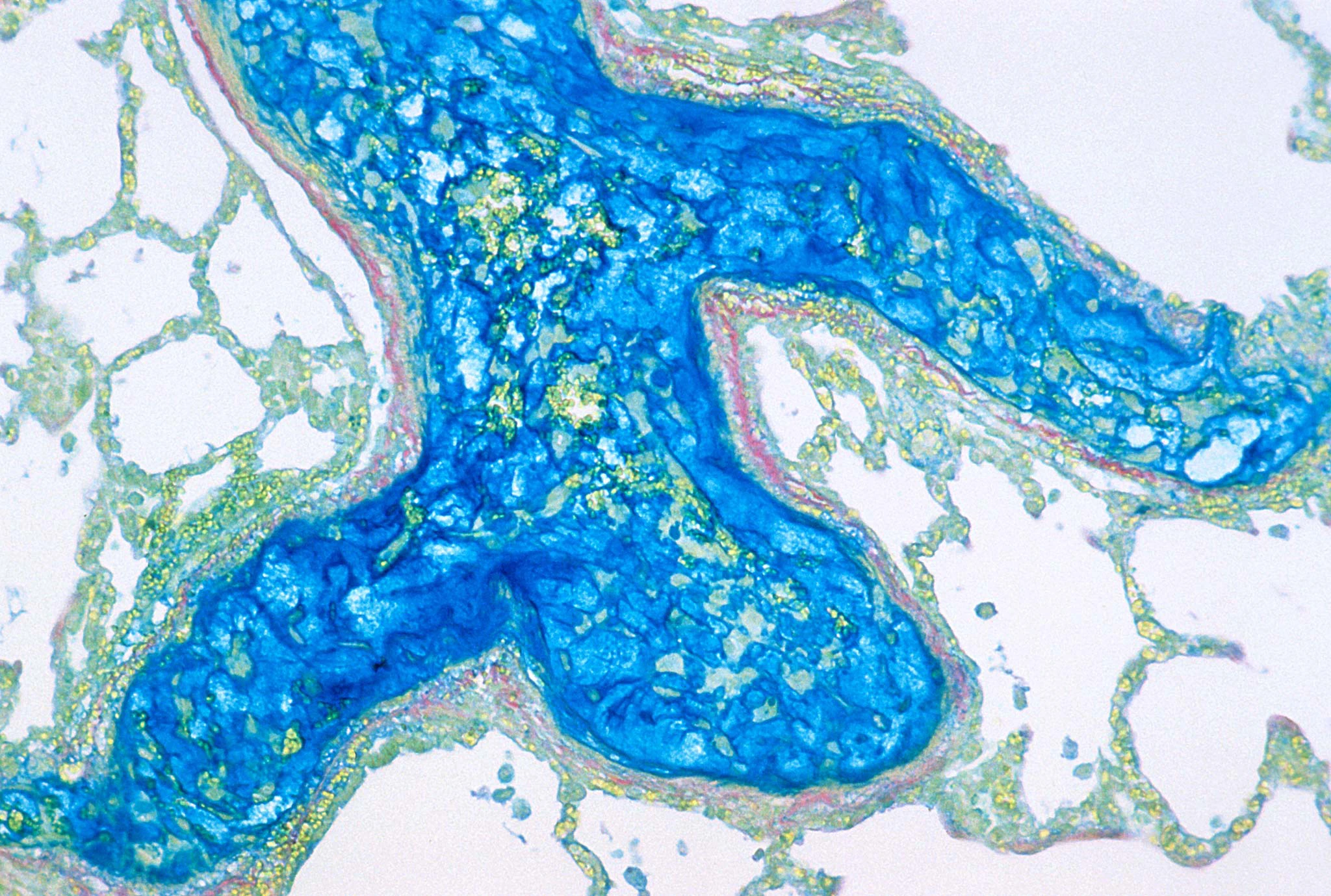
Image of metastatic pulmonary blastoma captured using a Nikon microscope
Enhancing diagnostic efficiency through digitization and digital slide scanning
In line with the digital transformation in healthcare, Nikon offers a complete solution for managing digitized slide images through an integrated digital slide scanning system featuring high-performance cameras and NIS-Elements software.
Nikon microscopes, combined with dedicated cameras, enable whole-slide imaging with automatic focus synchronization, high-resolution panorama stitching, and DICOM-compliant storage — supporting long-term archiving, remote consultation, and efficient image sharing.
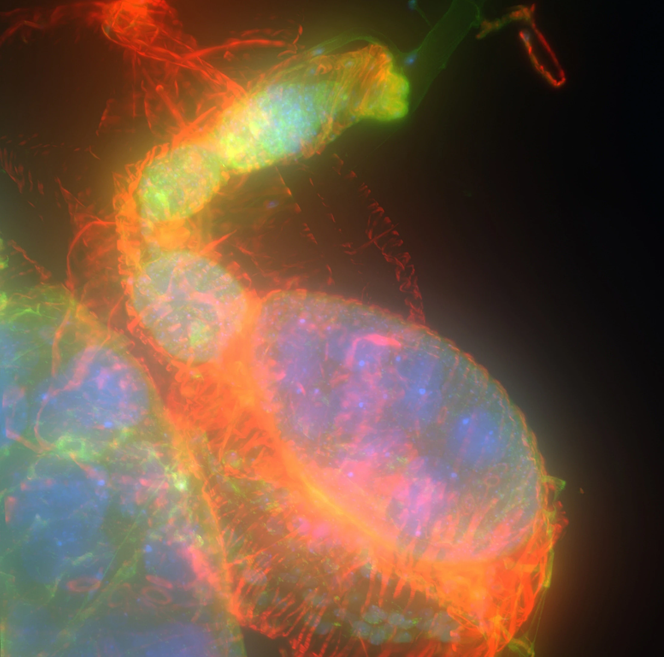
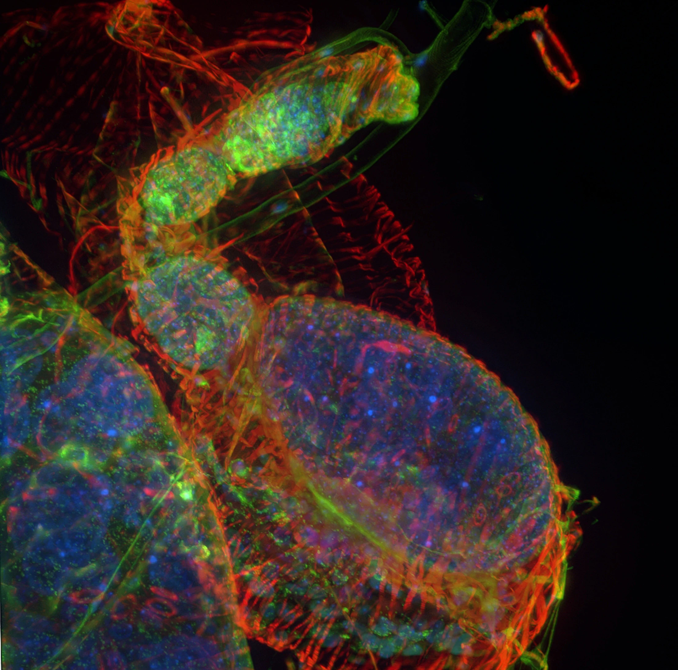
Image enhancement using AI tools
Fuorescence images before (left) and after (right) AI-based enhancement
NIS-Elements offers Z-stack capture, multi-channel fluorescence analysis, time-lapse imaging, macro documentation, quantitative reporting, and centralized image management — all enhanced by AI-powered processing and image optimization.
Microscope design optimized for long-term operation: wide field of view, flexible objective system, durable LED illumination, smooth stage movement, and seamless integration with motorized stages and AI-driven systems
Key Nikon Microscope Series Commonly Used in Pathology
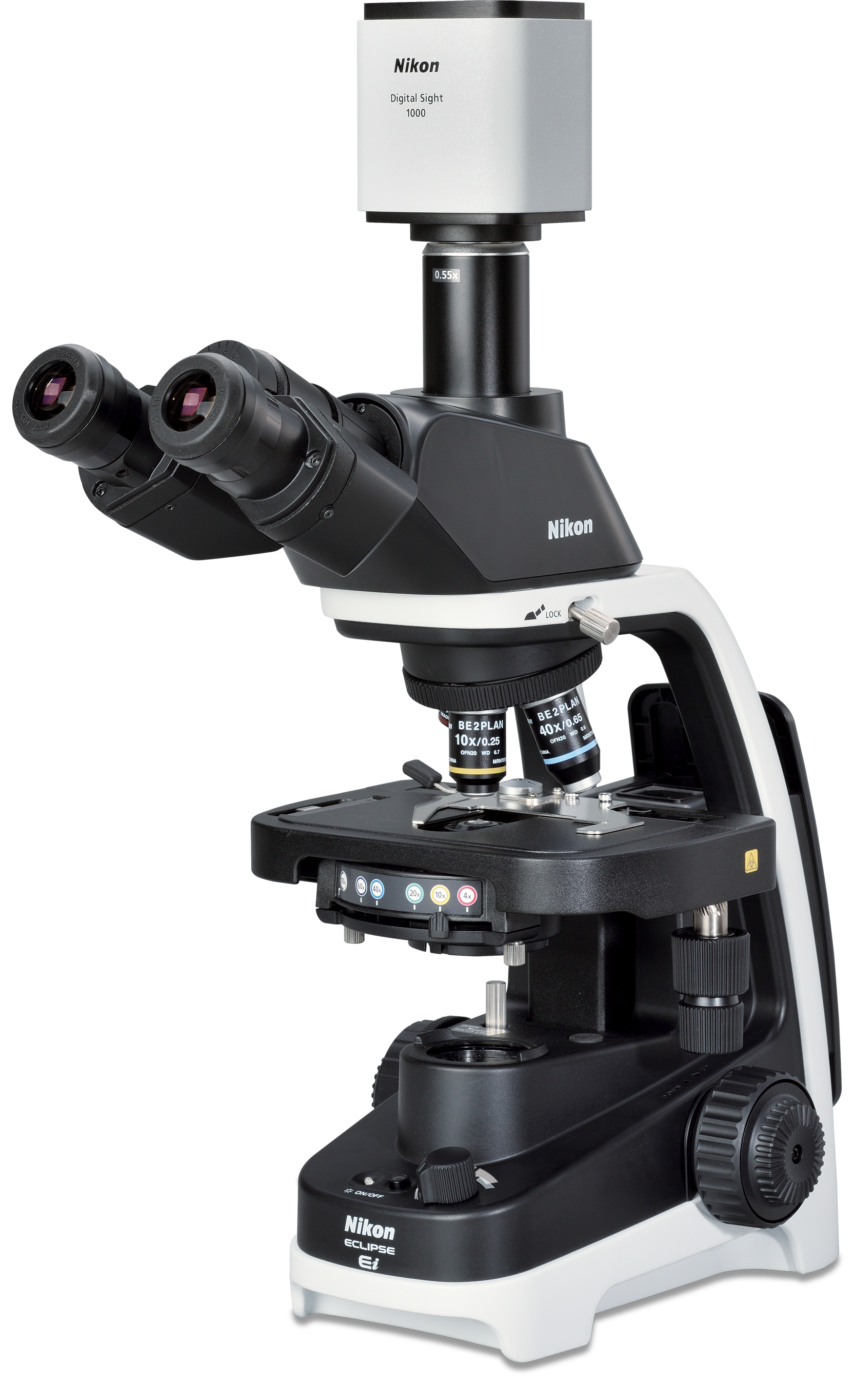
ECLIPSE Ei For Basic Observation
The 10X eyepiece and specialized CFI BE2 Plan Achromat objective system provide a wide field of view of up to 20 mm
Optional microscope cameras enable specimen image capture and real-time sharing via monitor or network.
A simple, compact design that saves space and is easy to transport.
Observation Method: Brightfield.
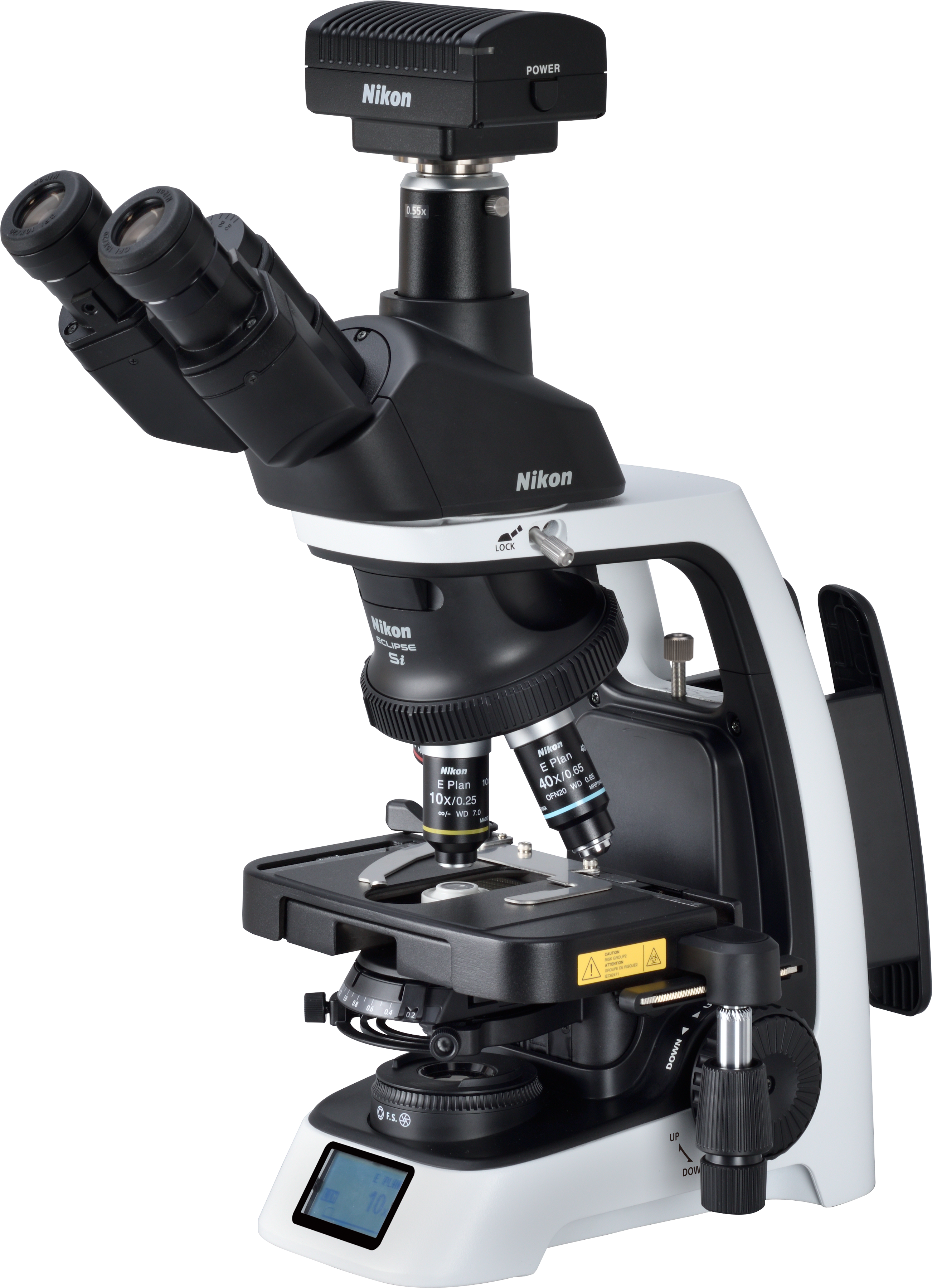
ECLIPSE Si:
Ergonomic design for long-lasting comfort in extended workflows.
LIM automatically stores and restores the appropriate light intensity for each objective.
Stage height limiter protects slides and objectives during focusing and sample changes.
Equipped with an LCD display showing magnification and light intensity
Observation methods can be switched using body-mounted control buttons; settings are automatically optimized based on the selected magnification on the Ni-E system.
Multiple motorized modules can be added to upgrade the system into a fully automated platform.
An integrated capture button ensures easy operation, while angled control buttons enable convenient touch-style image capture during observation.
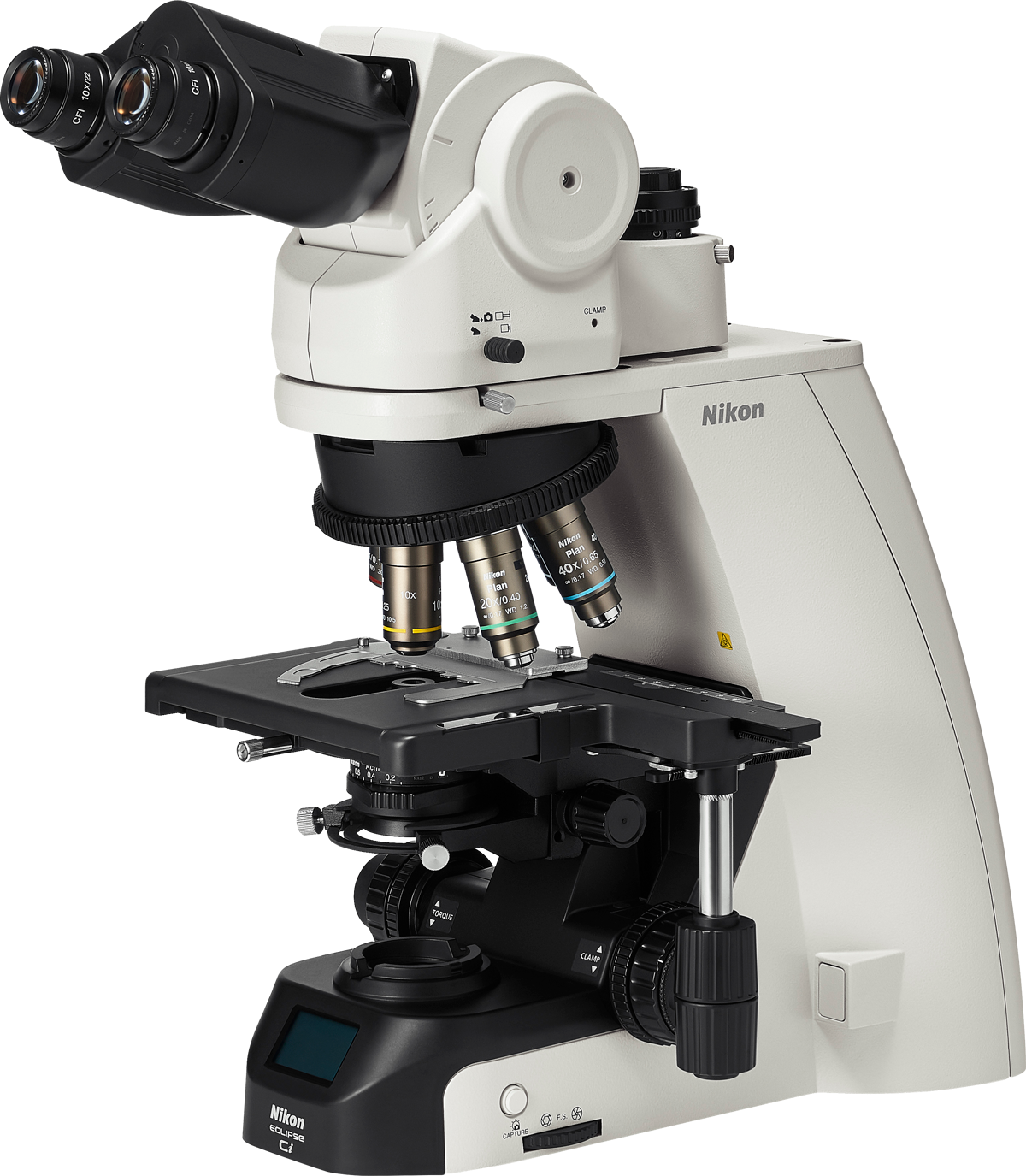
High optical performance is achieved through uniform, bright illumination with the Fly-eye optical system.
Optional LED light sources are available for brightfield and phase contrast observations
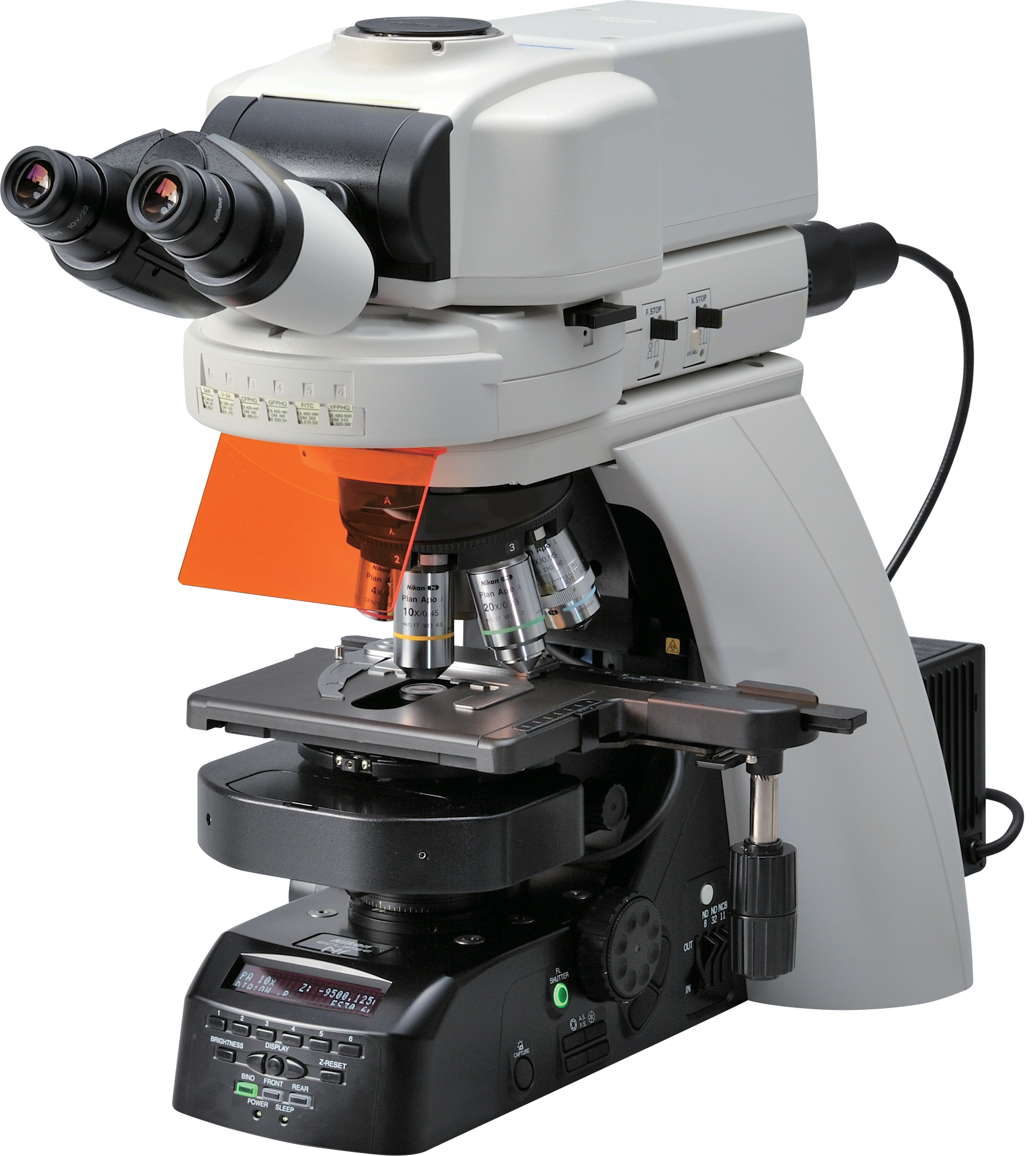
Supports up to 6 fluorescence channels and 7 objective lenses, enabling highly flexible imaging and application versatility.
More than just an instrument, Nikon microscopes are trusted partners in pathology. With outstanding optical quality, flexible integration, and digital pathology compatibility, Nikon is the ideal choice for modern pathology laboratories.