OUTSTANDING OPTICAL SOLUTIONS FOR CYTOLOGICAL AND HEMATOLOGICAL DIAGNOSTICS
In hematology and cytology, where every microscopic detail matters, Nikon microscopes deliver unmatched clarity, accurate color reproduction, and versatile design—making them the trusted choice in leading medical and research centers worldwide.
Widely and flexibly applied for accurate diagnostics
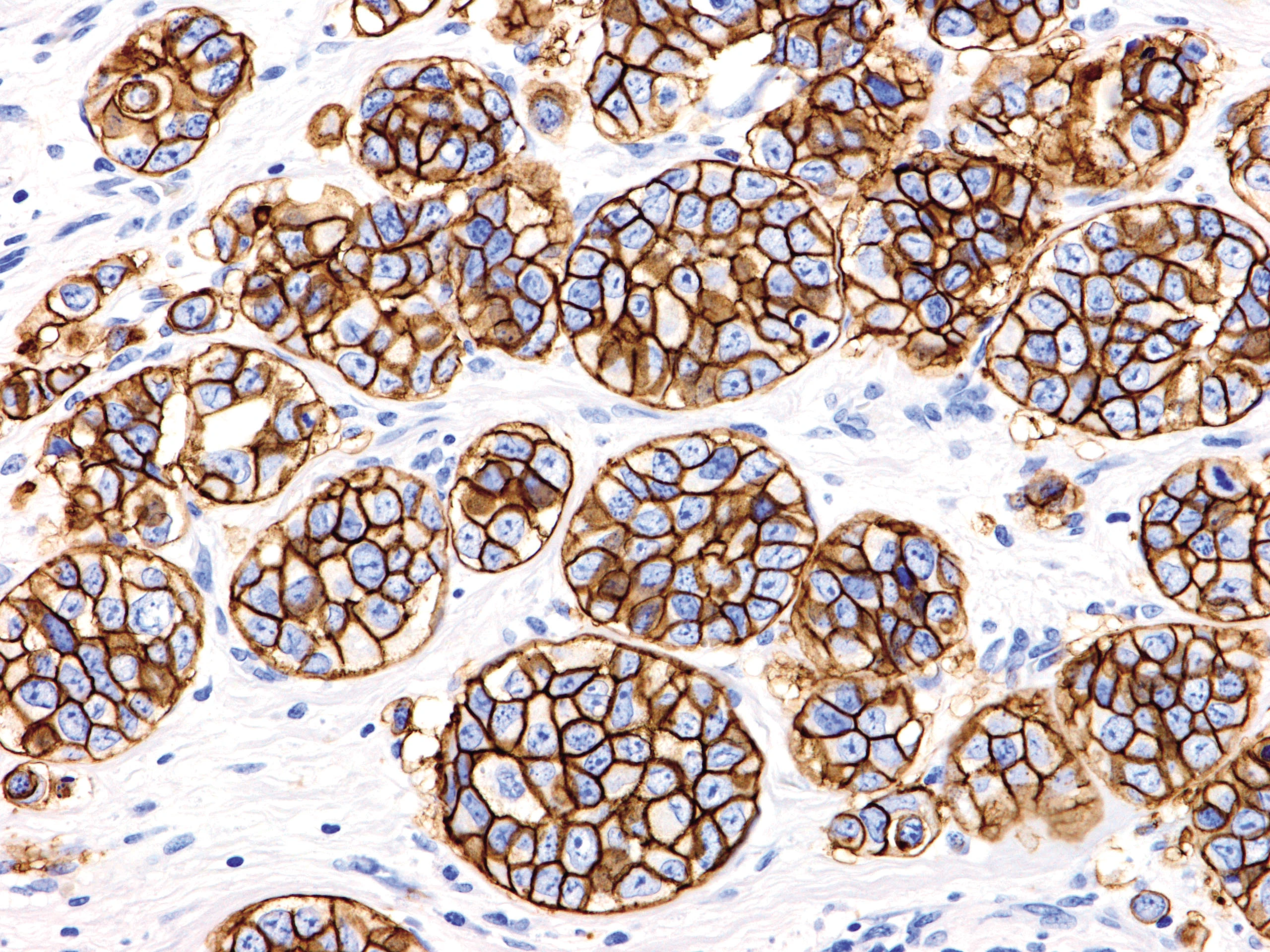
Nikon microscopes excel in peripheral blood smear analysis, accurately identifying white blood cell differentials, blast cells, and morphological changes. With true-to-life Wright-Giemsa color reproduction, Nikon is your trusted partner for reliable hematology diagnostics.
Tissue and cell sample images captured with Nikon microscopes
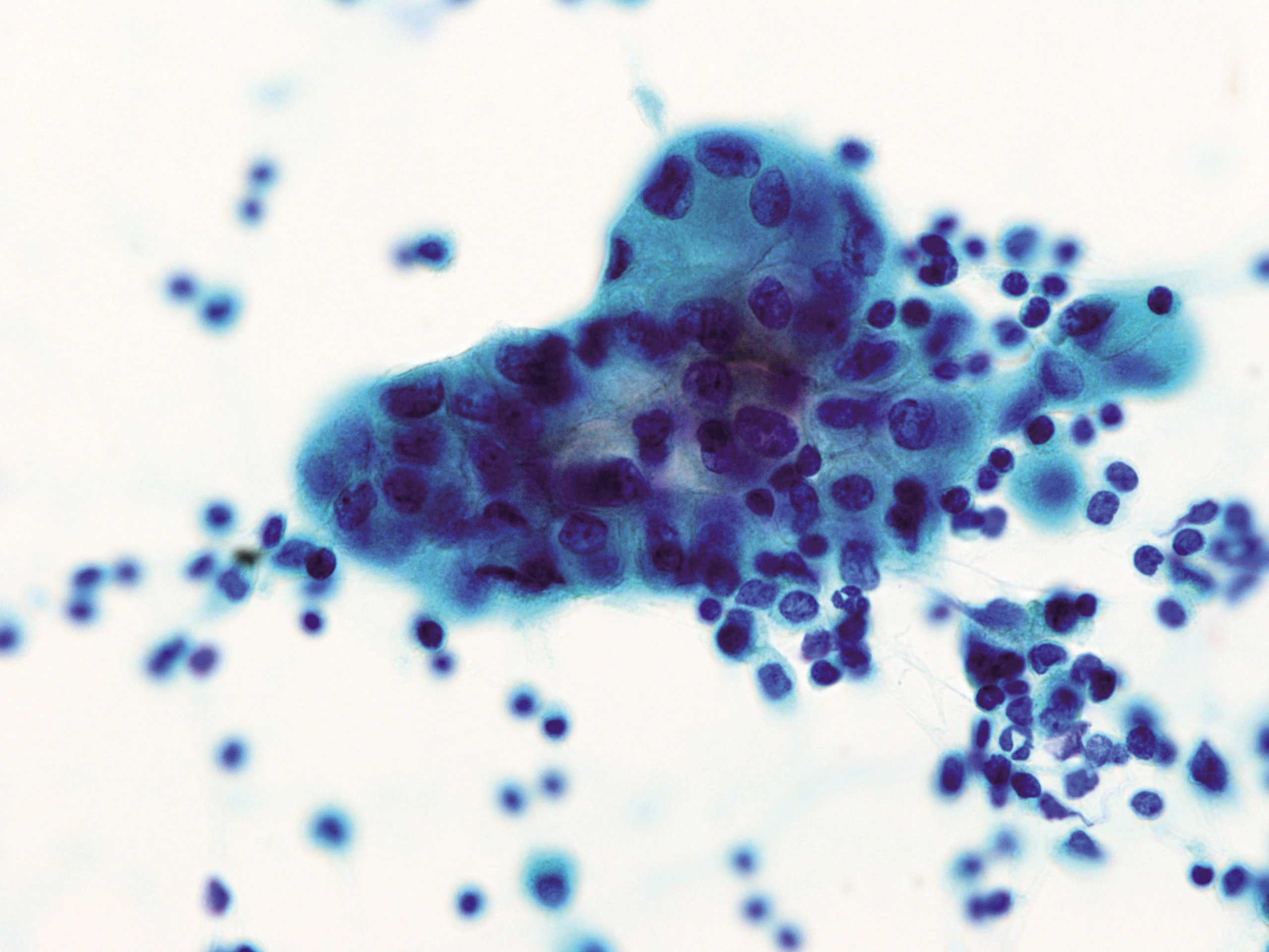
In cytology, Nikon microscopes are widely used for cervical smear analysis following the Bethesda system, as well as for examining fluid samples and fine-needle aspirations (FNA). With Nikon microscopes, you can clearly observe critical morphological features such as hyperchromatic nuclei, dysplasia, and abnormal mitoses—supporting clinicians in making accurate assessments of lesion nature.
Beyond that, Nikon microscopes play an essential role in cultured cell analysis, drug response evaluation, cell density and cycle measurement, as well as apoptosis identification through nuclear morphology. When combined with digital cameras and NIS-Elements software, the system supports slide digitization, semi-automated measurements, statistical analysis, and data storage to facilitate research and scientific publication..
Quality built on a foundation of precision optics and accurate microscopic analysis
The core technology of Nikon microscopes is the CFI60 infinity optical system, delivering high precision, wide field of view, and optimal light transmission. This system is especially suited for observing thin cytology specimens rich in morphological details, such as PAP smears and fine-needle aspirations (FNA).
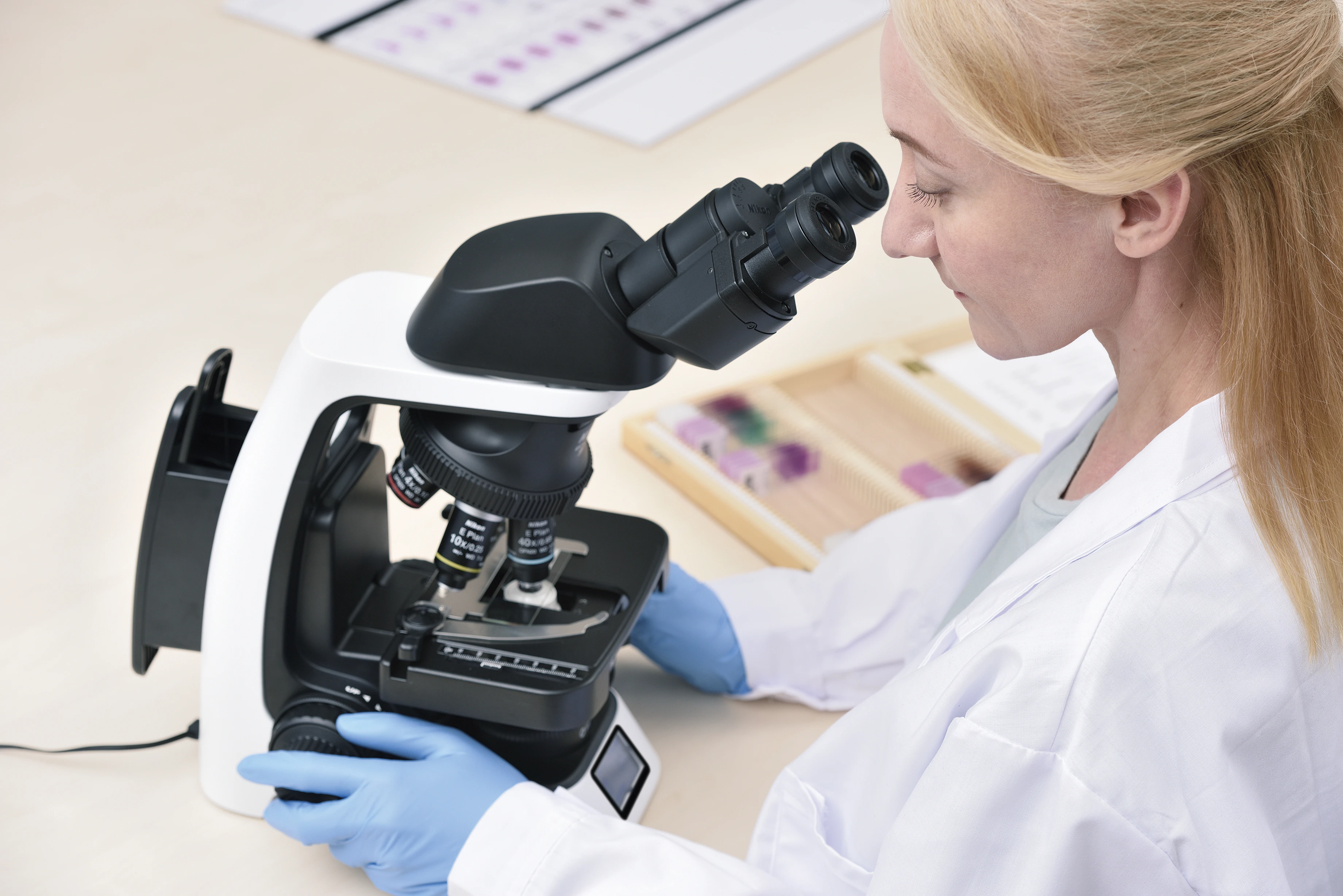
With an adjustable condenser, durable LED illumination, objective lenses ranging from 4x to 100x, and a user-friendly design, Nikon microscopes effectively meet both routine diagnostic and advanced analytical needs. Models such as the Eclipse Ci-L, Ni-U, and Ni-E offer flexible customization with a wide range of specialized accessories, making them suitable for diverse applications.
Integrated Digital Technology – A Leap Forward in Healthcare Digital Transformation
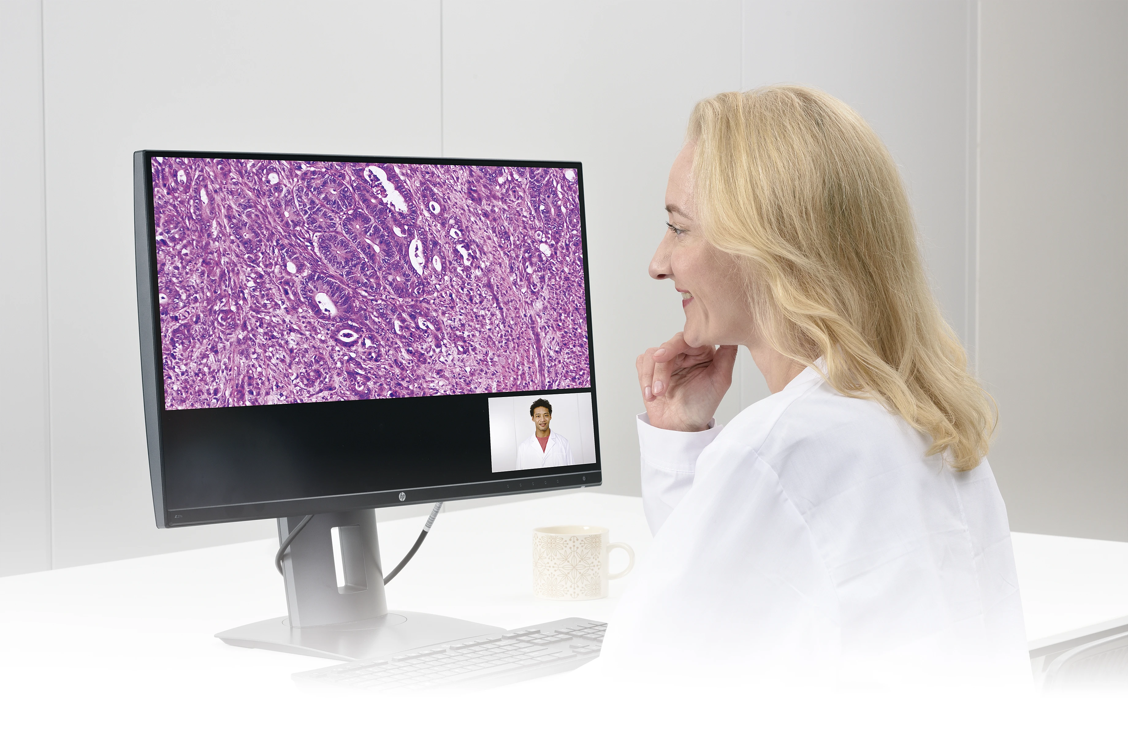
Nikon microscopes serve as a powerful platform for digital pathology, enabling high-resolution slide imaging, clinical data tagging, and seamless integration with PACS/LIS systems. More than just an optical tool, Nikon combines precision optics, ergonomic design, and digital readiness to support accurate diagnosis, case archiving, teleconsultation, and AI-driven applications.
With outstanding color fidelity, broad objective range, and flexible configuration—from routine screening to advanced analysis—Nikon is the strategic choice for modern laboratories aiming toward diagnostic excellence and digital innovation.