Chromosome analysis plays a vital role in detecting genetic abnormalities associated with congenital disorders, developmental anomalies, and cancer.
Two core techniques widely applied in cytogenetics are karyotyping and fluorescence in situ hybridization (FISH).
While karyotyping enables comprehensive visualization of the entire chromosome set—identifying major numerical and structural abnormalities—FISH allows for the detection of microscopic alterations with high specificity.
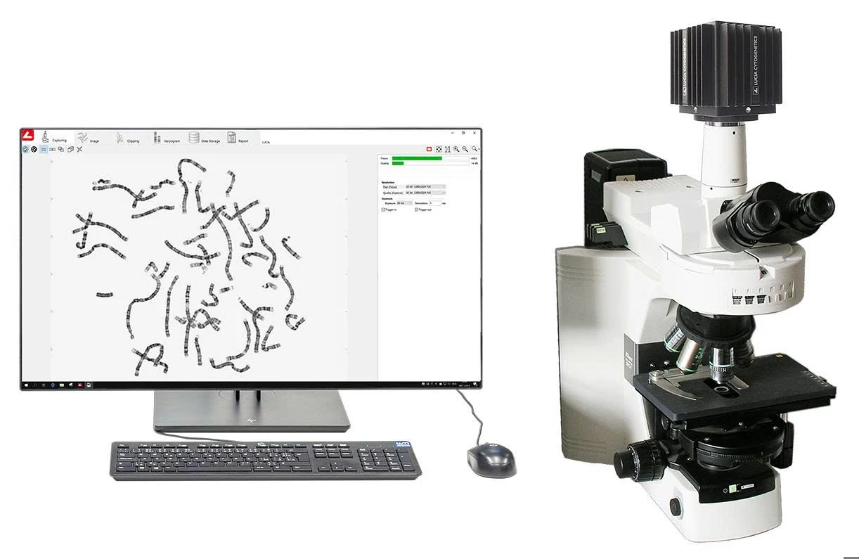
Karyotyping with Lucia Cytogenetics
Karyotyping:
Karyotyping is a chromosome morphology analysis technique based on characteristic banding patterns. It is typically performed during the metaphase stage of mitosis, when chromosomes are maximally condensed for clear visualization. The most widely used method is G-banding (Giemsa banding), which enables the identification of individual chromosomes by their distinct light and dark banding patterns.
The standard procedure includes several key steps: collecting cell samples (such as peripheral blood, amniotic fluid, chorionic villi, or bone marrow), culturing them in specific media, treating with colchicine to arrest cells in metaphase, harvesting, fixing, and spreading the cells onto microscope slides. After staining, chromosomes are analyzed under a light microscope, imaged, and arranged into a standardized karyotype.
The Nikon microscope system integrated with Karyo software supports you with:
Slide image scanning: capturing large-scale images and seamless image stitching
Automatic camera parameter settings for optimized image capture
Chromosome image acquisition after cell membrane lysis
Assistance in chromosome separation and isolation
Automated chromosome classification and karyogram generation
Customizable karyogram layout tools
Image editing and enhancement tools for chromosome visualization
Intuitive chromosome comparison tools
Action history tracking for editing and analysis steps
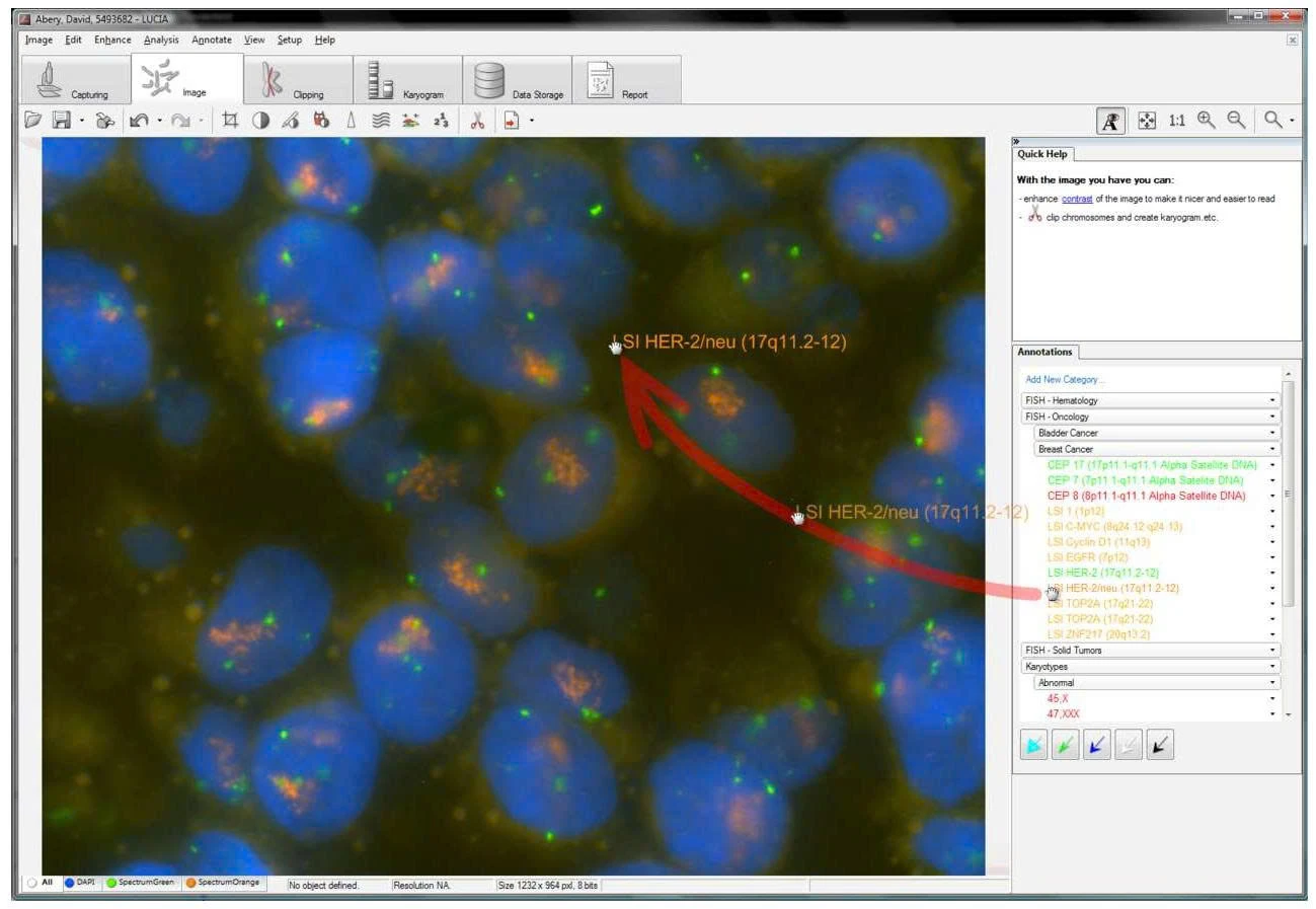
FISH: Mechanism and Broad Applications
Fluorescence In Situ Hybridization (FISH) is a modern technique that uses fluorescently labeled, sequence-specific DNA probes to hybridize with target DNA sequences within cells. The hybridization occurs directly on fixed slides, allowing precise localization of specific genes or chromosomal regions of interest.
FISH software
The key advantages of FISH include its ability to detect very small abnormalities (less than 1 Mb), high specificity, rapid turnaround time, and applicability to both dividing (metaphase) and non-dividing (interphase) cells. This makes it particularly valuable in urgent situations such as rapid prenatal diagnosis, analysis of paraffin-embedded tumor tissues, or detection of specific translocations in hematologic malignancies.
Unlock accurate and efficient fluorescence analysis with Nikon’s integrated microscope and FISH software system. Key features include:
Smart control of microscope and camera
Optimized imaging presets for each probe set
Intelligent image acquisition with auto parameter adjustment
Extended Depth of Focus (EDF) for thick specimen
Automatic probe-to-layer assignment with metadata tracking
Accurate color validation across image components
Built-in tools for image enhancement and annotation
Karyotyping and FISH are essential tools in cytogenetic diagnosis. Karyotyping provides a broad view of chromosomes, while FISH offers precise, rapid detection of microscopic abnormalities.
To maximize these techniques, effective optical systems and powerful software are crucial. SISC proudly offers an integrated solution combining Nikon microscopes with LUCIA software, supporting clinical and research needs in prenatal medicine, hematology, oncology, and reproductive health.
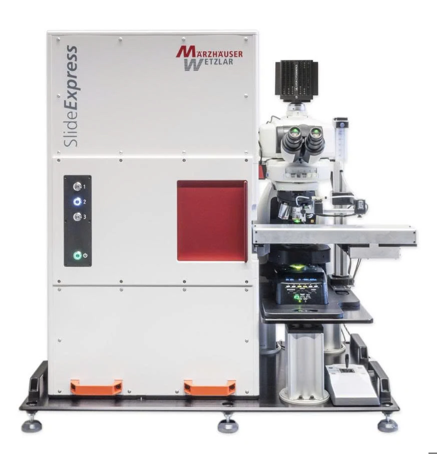
ECLIPSE Ni-E and SlideExpress System
Nikon microscopes feature the CFI60 Infinity Optical System, enhancing contrast, flatness, and resolution with a wide 25 mm field of view for detailed image capture. For karyotyping, they provide sharp metaphase chromosome images at high magnification (usually 100x oil objective) for G-banding and karyotype analysis. In FISH, they ensure high sensitivity to weak fluorescence signals and clear color separation with specialized filter cubes and stable illumination.
The Nikon Eclipse Ni-E automated microscope features precise autofocus, motorized objective turret, and motorized stage movement. These capabilities provide a foundation for automated metaphase search and fully automated slide scanning.
Lucia Metaphase Finder with Märzhäuser SlideExpress 2
Alongside the optical hardware, LUCIA Cytogenetics software (developed by Laboratory Imaging, Czech Republic) serves as a specialized image analysis platform for cytogenetic applications. LUCIA software suites are powerful laboratory tools, offering advanced features to:
Automatic metaphase search and chromosome image acquisition.
Automated or semi-automated karyotype arrangement according to ISCN standards.
Chromosome naming, numbering, measurement of length, centromere ratio, and morphological parameters.
Detection of aneuploidy, translocations, deletions, and duplications in chromosome structure analysis.
Fluorescence signal analysis in FISH: signal counting, relative positioning, area and intensity measurement, differentiation of fusion, deletion, or gene amplification events.
Integration of case management modules, standardized report generation, and compliance with international data storage regulations (GLP/GMP).
The combination of Nikon microscopes, analysis software, and Slide Express offers a fully automated solution for karyotyping and FISH, capable of handling up to 120 slides per run. This system reduces repetitive work, allowing specialists to focus on advanced analysis.
Leveraging Nikon’s advanced optics and LUCIA’s powerful image processing, it supports prenatal genetic screening, malignant hematology diagnosis (e.g., CML, AML), and assisted reproduction research. Widely adopted by biomedical centers, hospitals, and research institutes in Vietnam and globally.