UNLOCK THE INVISIBLE – ADVANCED CYTOLOGY WITH NIKON MICROSCOPES
Through its dedicated solutions for cytological analysis, NIKON has established itself as the preferred strategic partner for thousands of laboratories, cellular research centers, oncology institutes, and regenerative medicine facilities across the globe—trusted for its precision, reliability, and innovation.
Nikon's Microscope Lineup for Cellular Research
APPLICATIONS OF NIKON MICROSCOPES IN BASIC RESEARCH
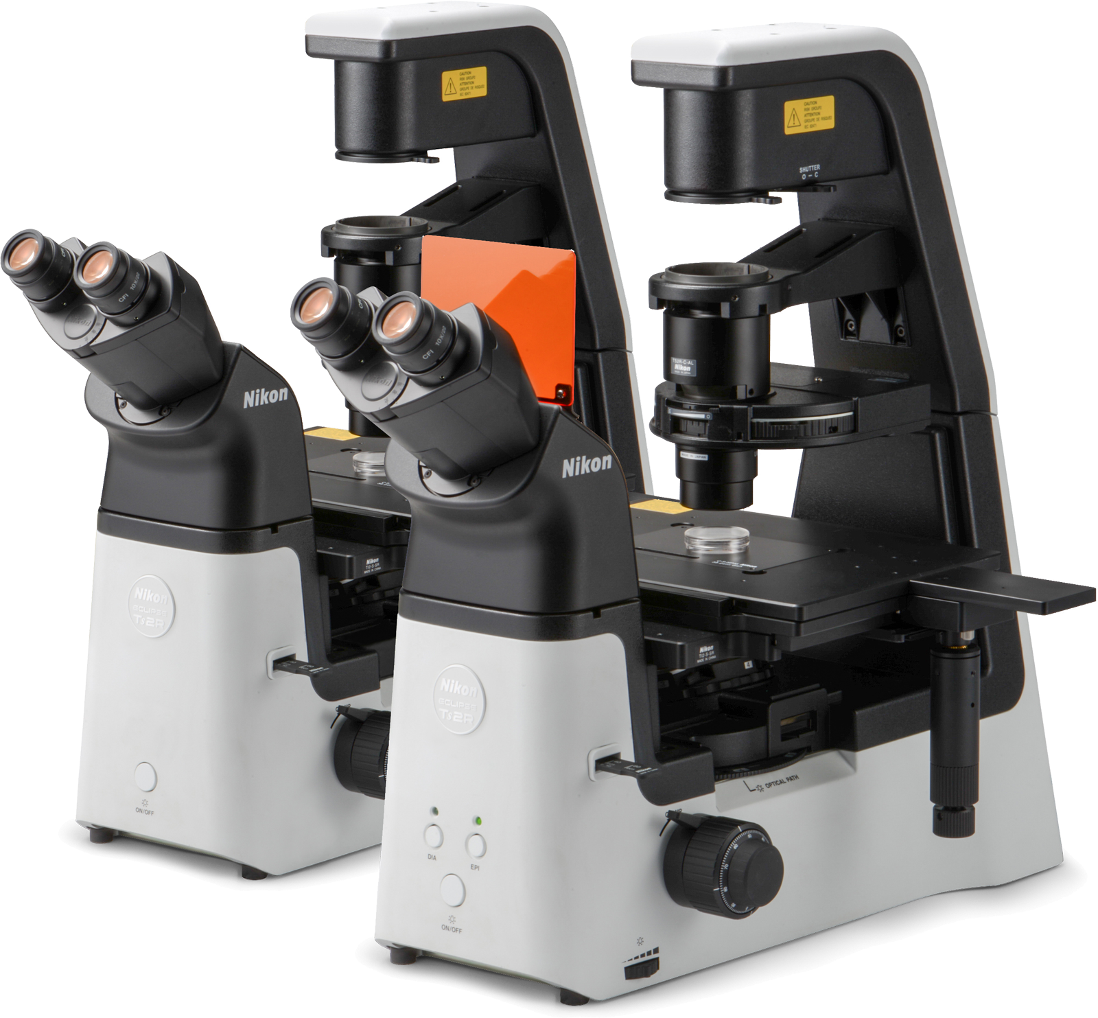
ECLIPSE Ts2/Ts2-FL
Nikon’s advanced CFI60 optics system, paired with a long-lasting 60,000-hour white LED and fly-eye lens technology, ensures evenly distributed, high-quality illumination throughout the entire field of view—ideal for both routine and high-precision microscopy
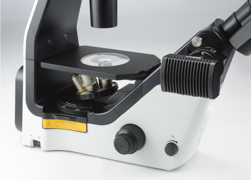
Simple and Efficient Design, Compact Size for Easy Installation
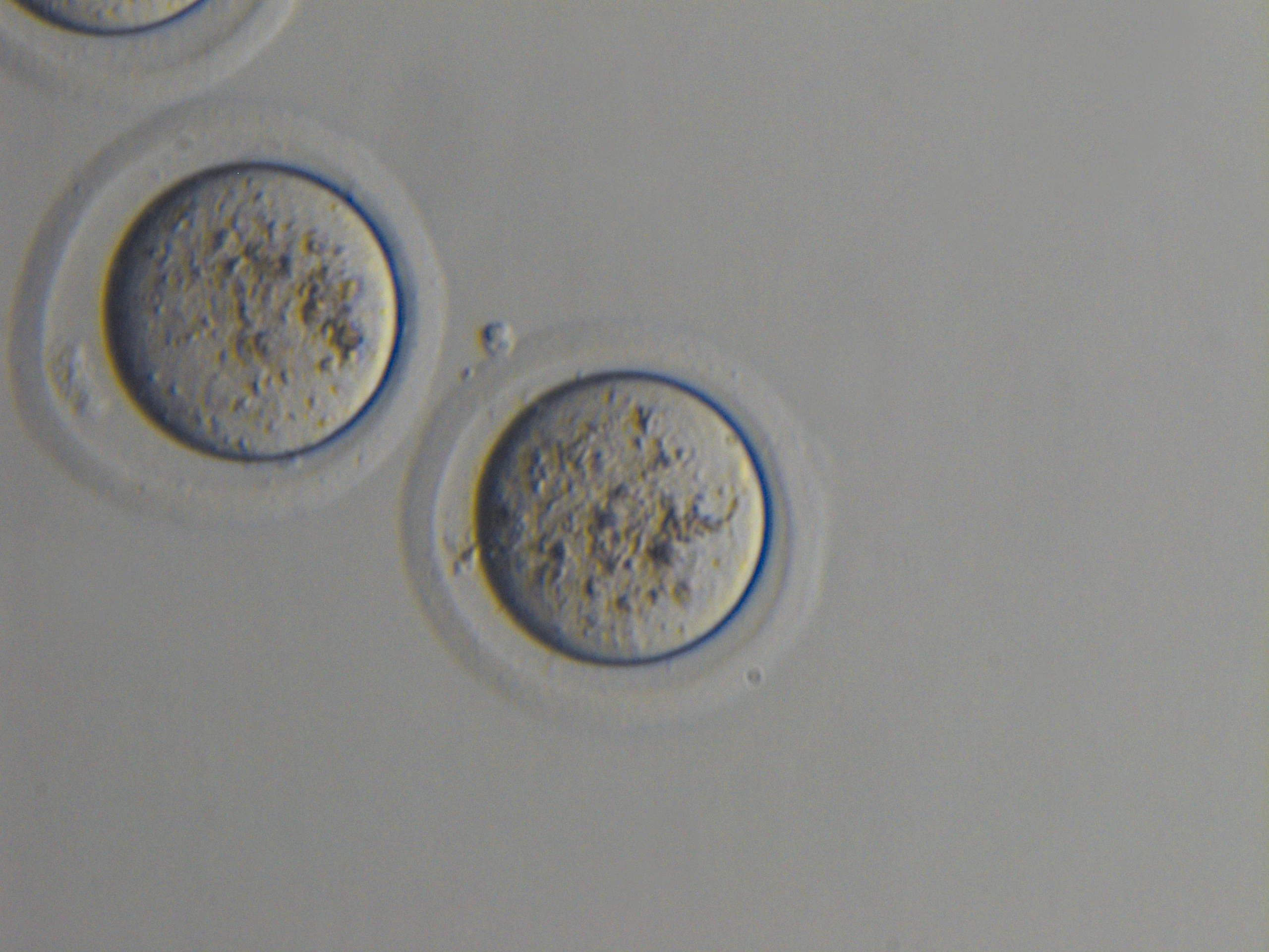
The Emboss Contrast technique creates a pseudo-3D effect with high-contrast imagery, enhancing depth perception in transparent specimens.
Quan sát huỳnh quang trên Ts2 FL
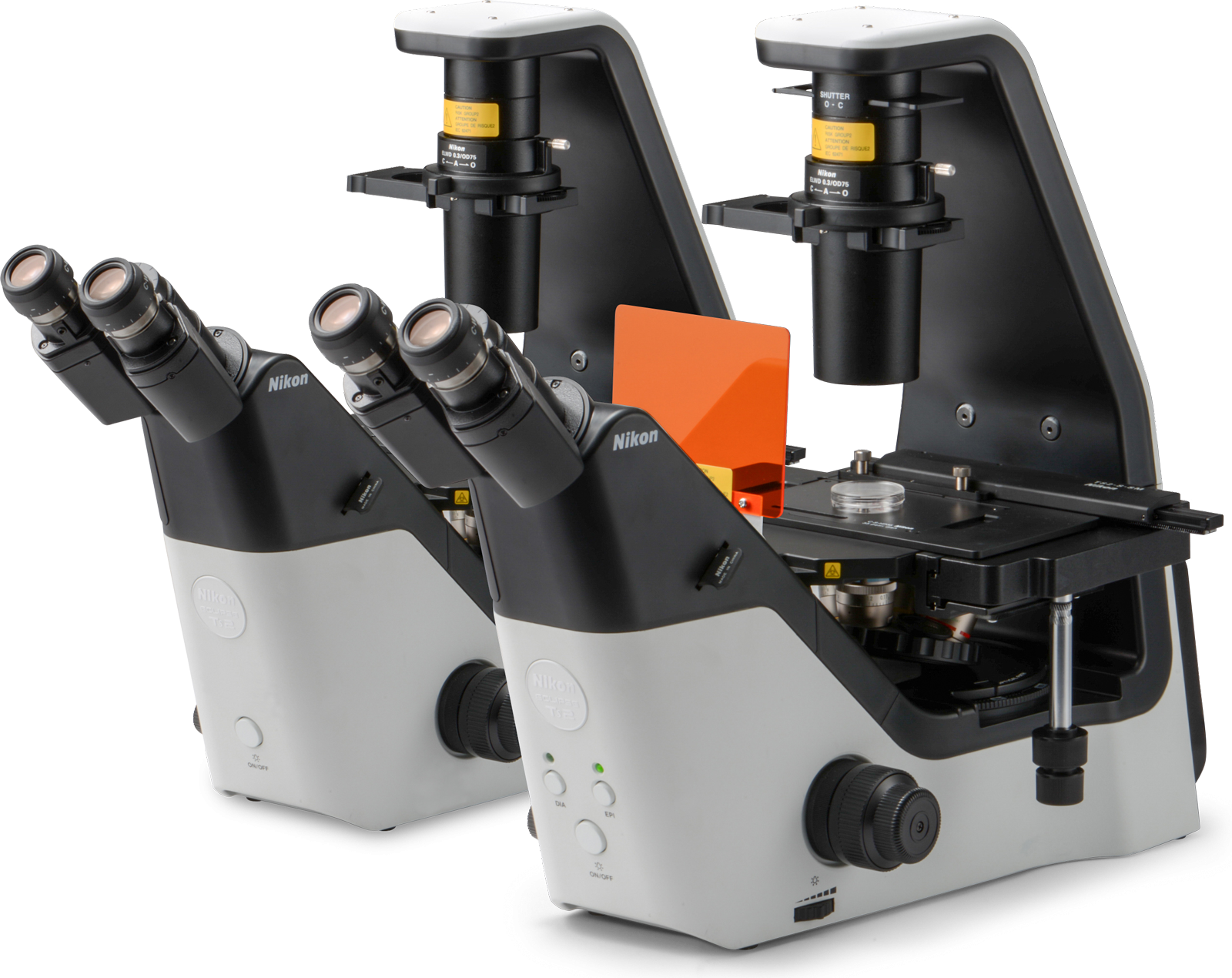
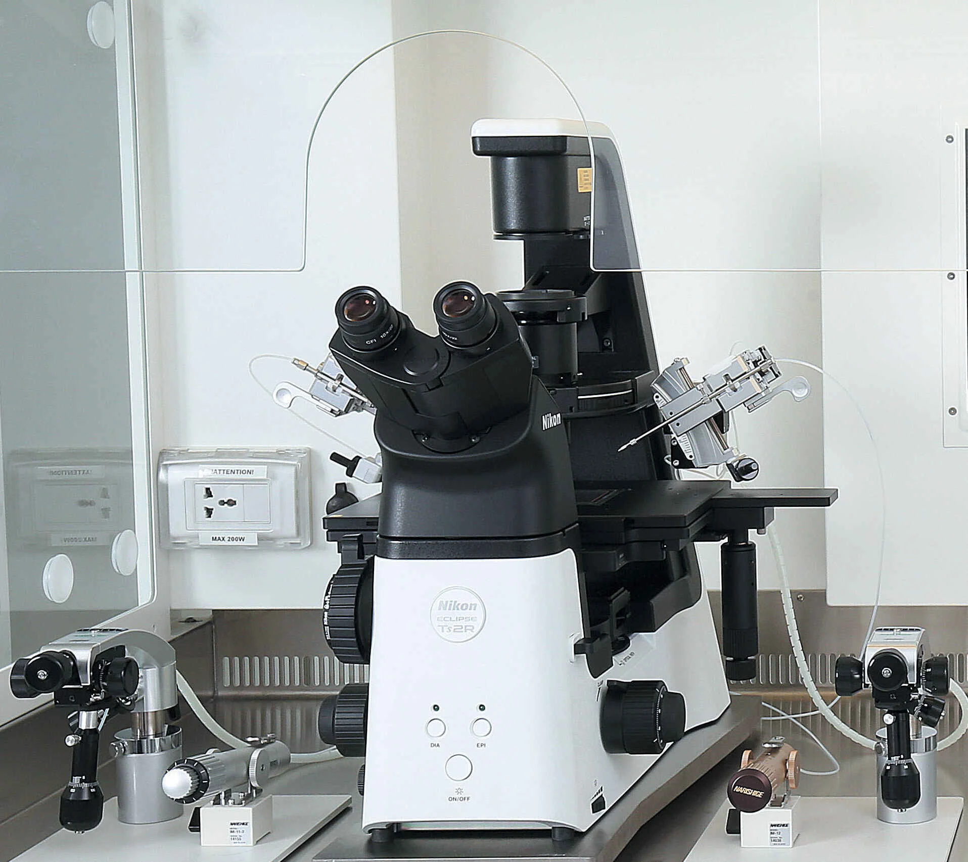
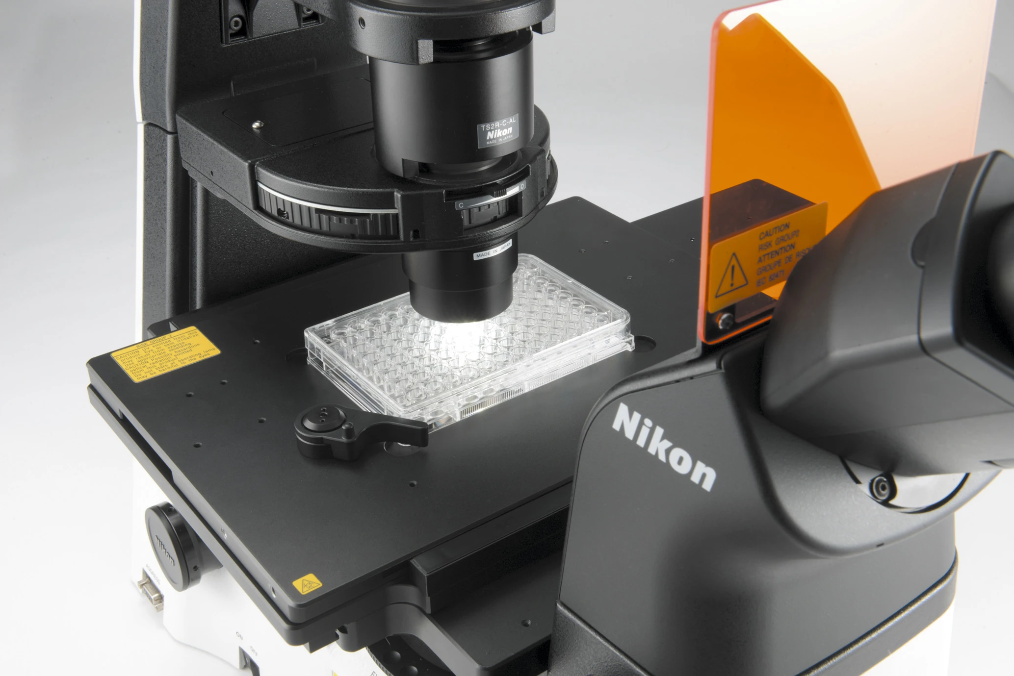
NIKON ECLIPSE Ts2R/Ts2R FL
Compact – Intuitive – Powerful, with High Ergonomic Performance.
Supports Advanced Observation Methods such as DIC and NAMC—on par with high-end research microscopes.
A large mechanical stage with extended travel range enables convenient observation of 96-well plates.
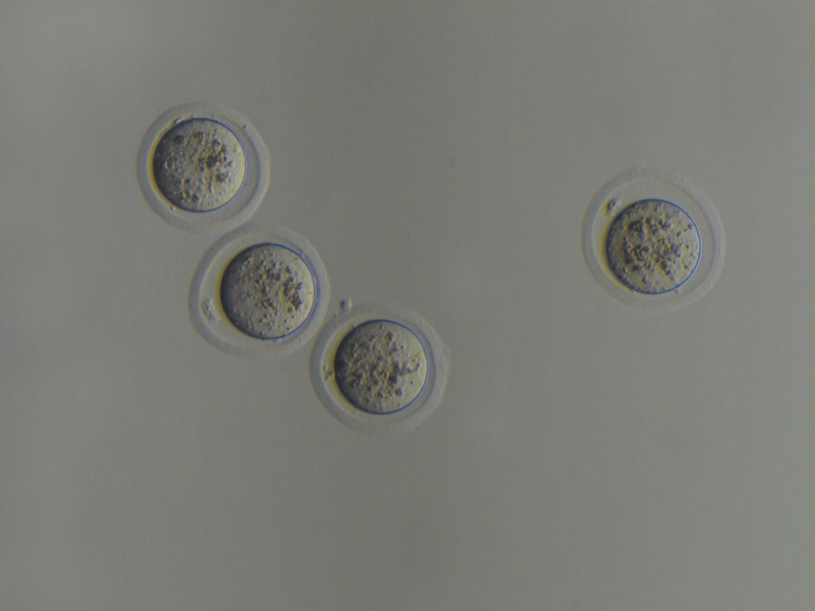
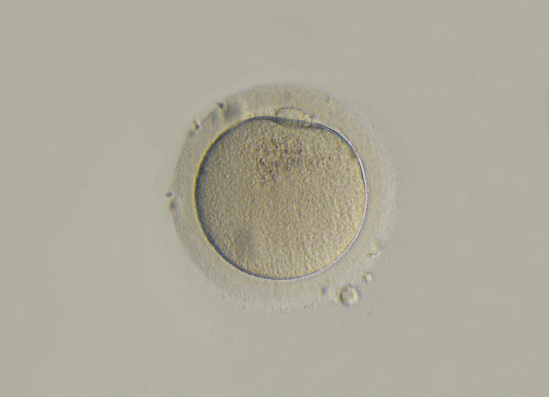
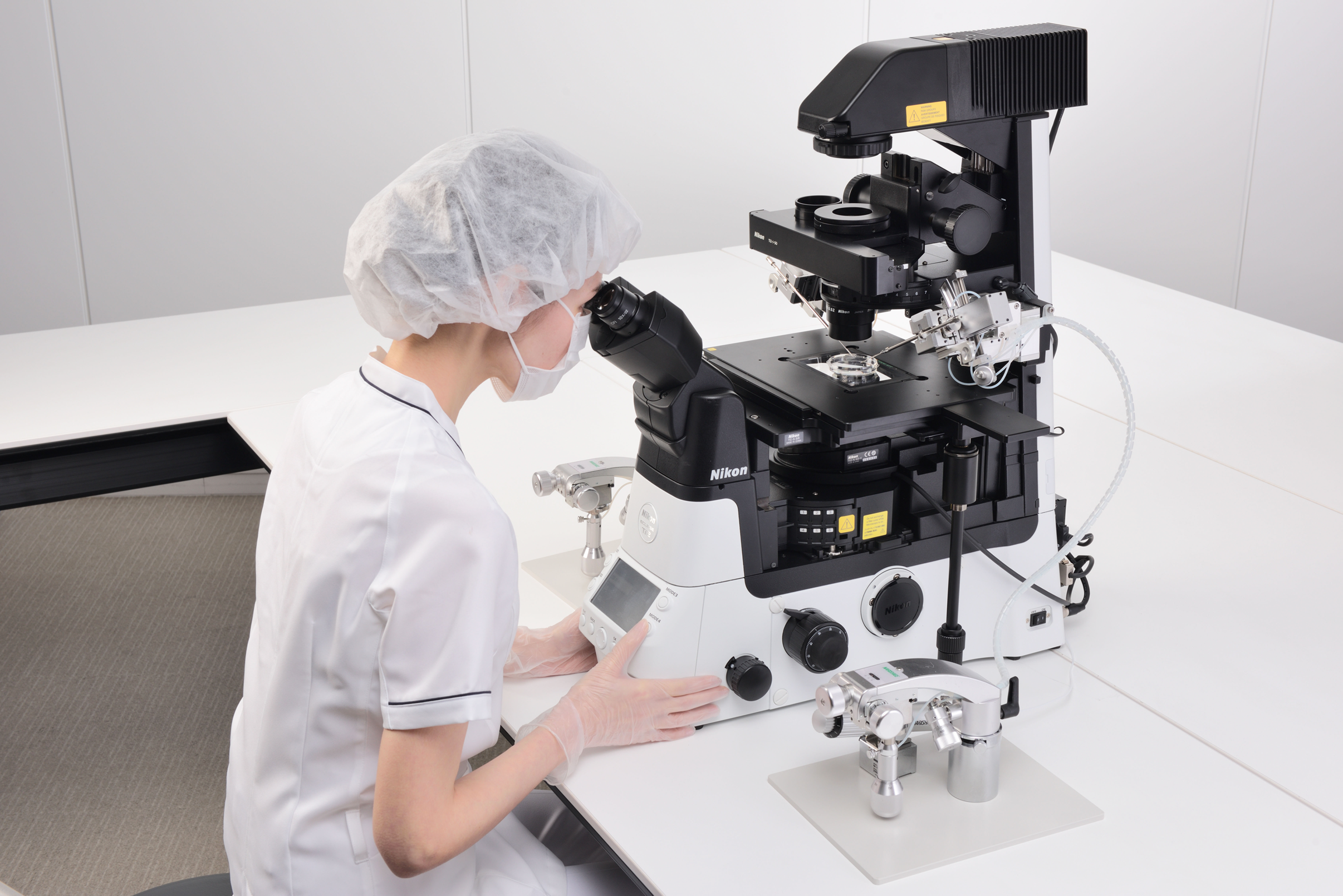
Compatible with Micromanipulator Systems for IVF Applications
Designed for high-throughput workflows, the spacious mechanical stage supports full 96-well plate scanning with ease. A 30% reduction in stage height enhances ergonomic comfort, minimizing strain during extended operation.
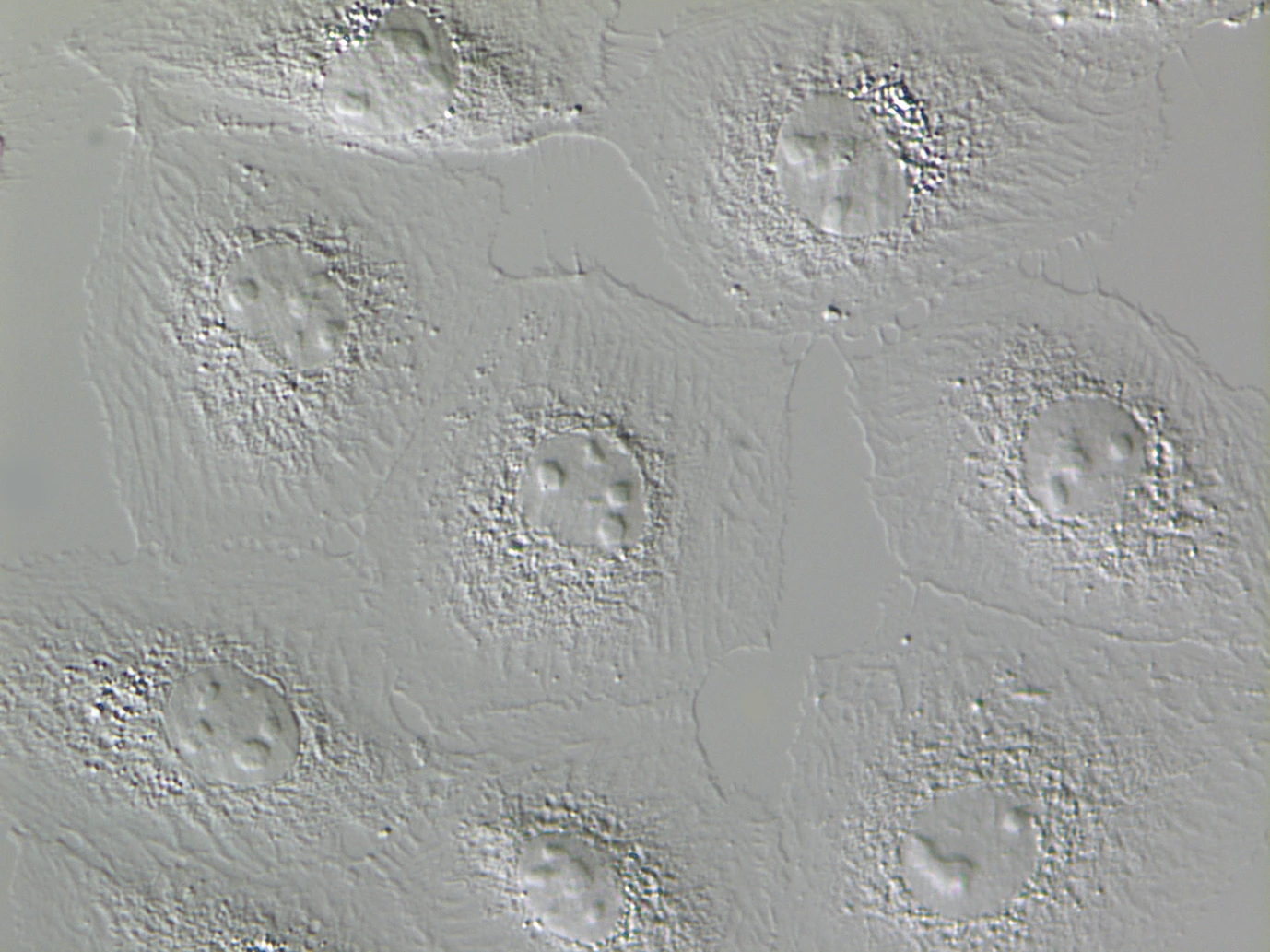
DIC and Emboss Contrast Imaging on the Eclipse Ts2R / Ts2R-FL
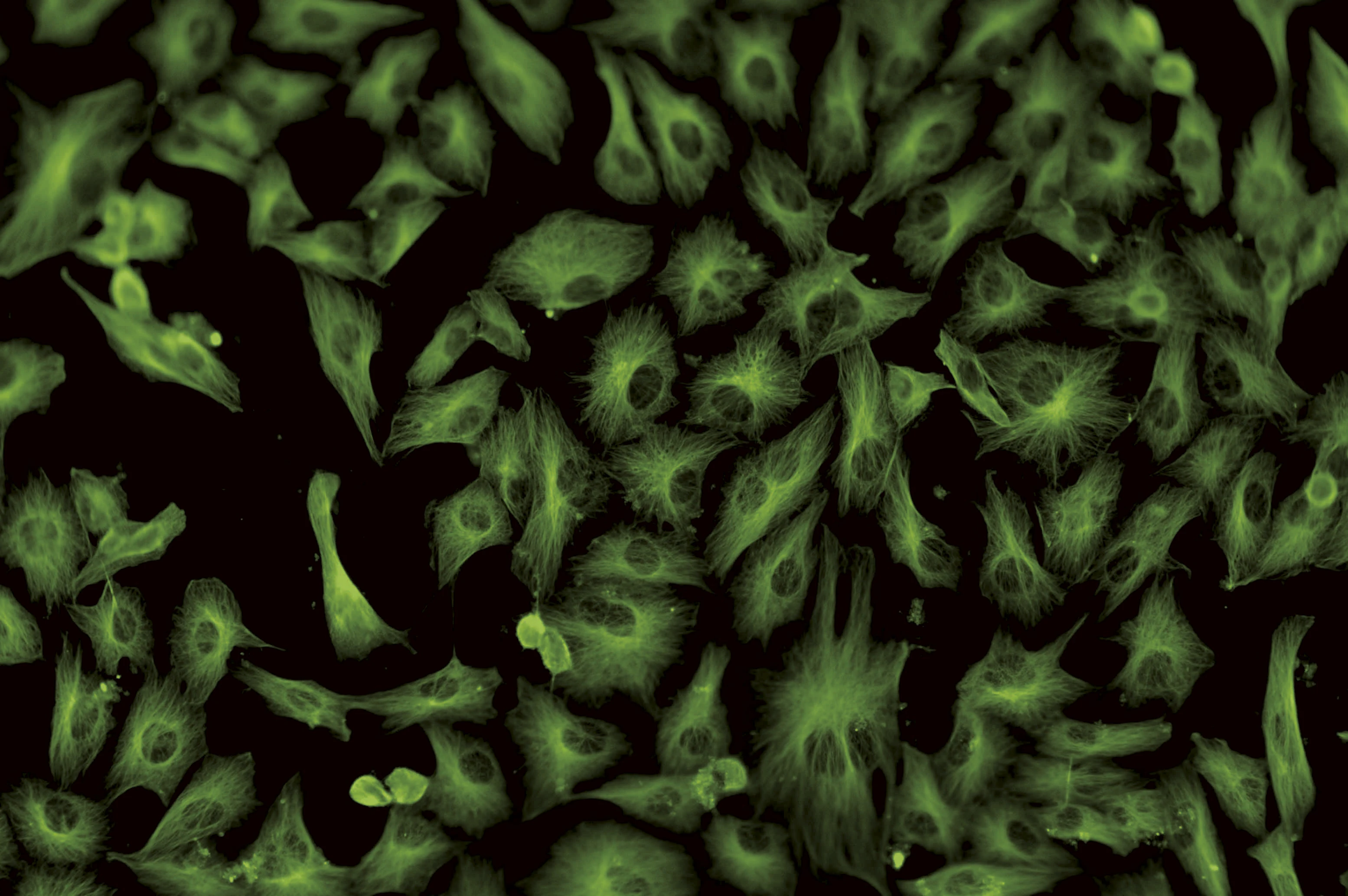
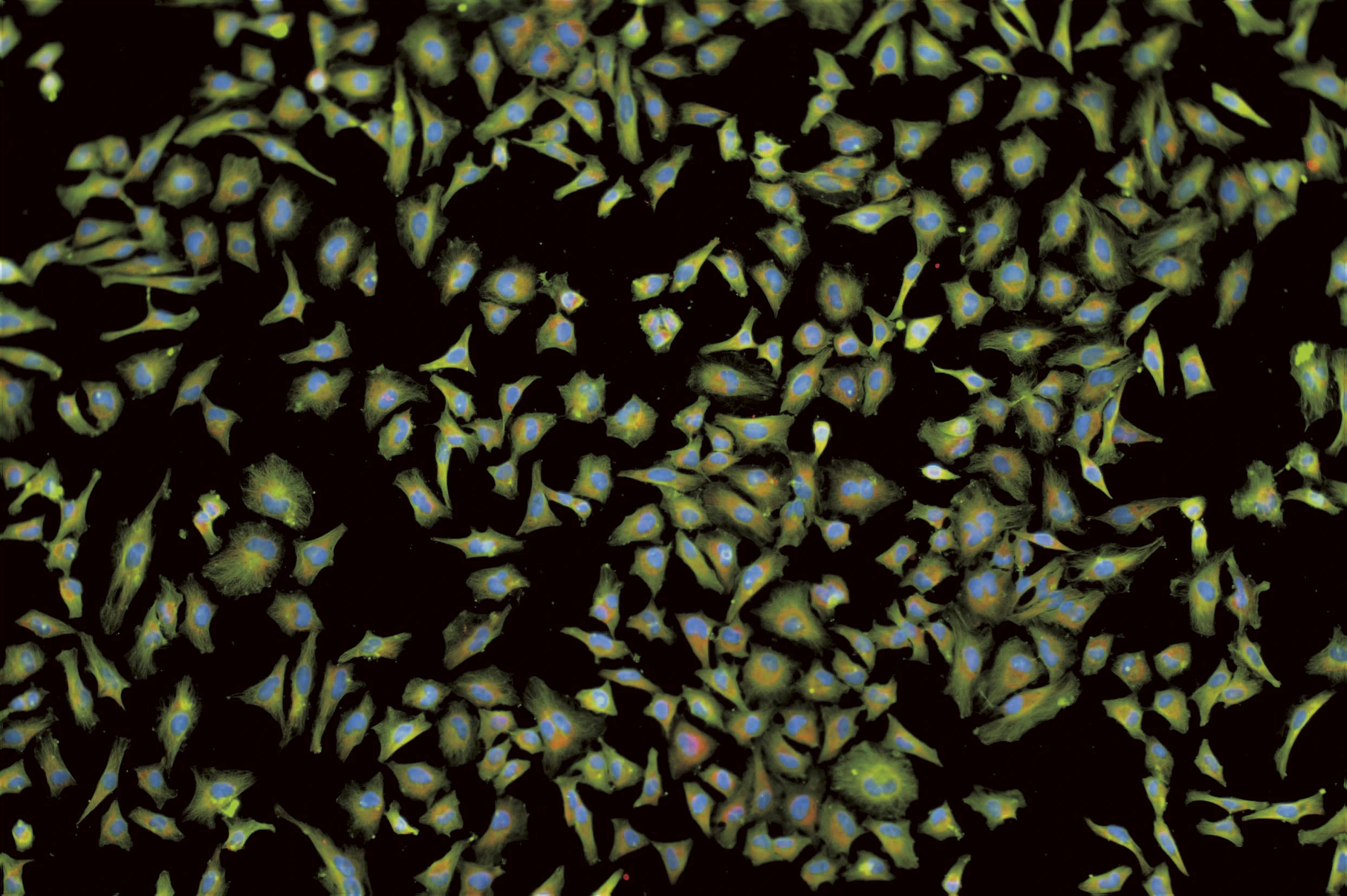
Fluorescence images captured on the Ts2R-FL, showcasing both single-channel and multi-color labeling for diverse cellular applications
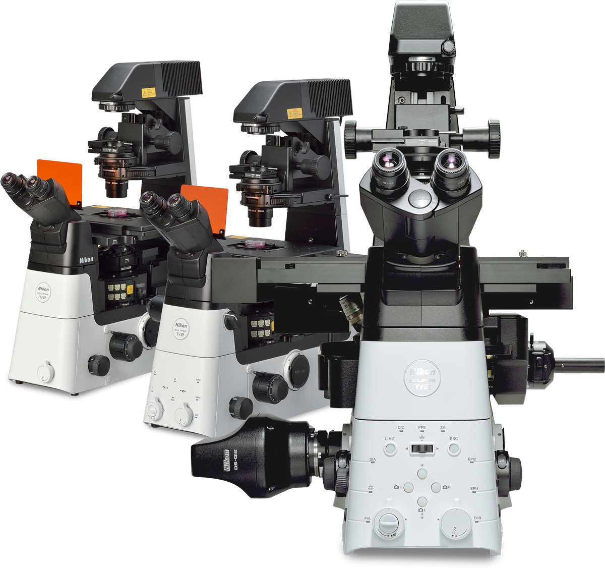
APPLICATIONS OF NIKON MICROSCOPES IN ADVANCED RESEARCH
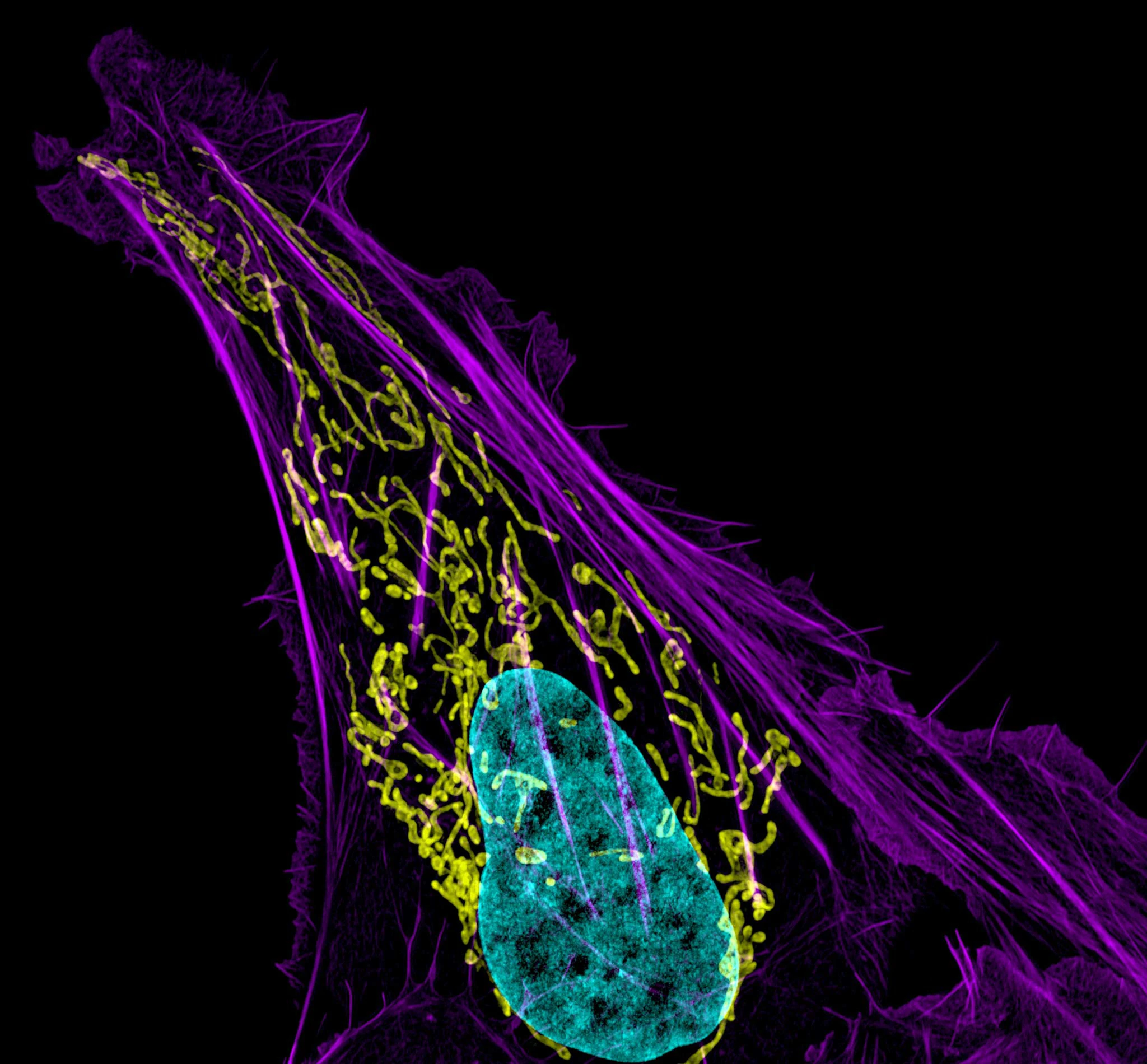
In cellular structural studies, Nikon microscopes provide high-resolution, aberration-corrected objectives for clear visualization of organelles such as the nucleus, nucleolus, ER, Golgi, and mitochondria in both live and fixed cells. These systems are essential tools for exploring the spatial distribution of proteins, DNA, RNA, and intracellular signals using multicolor fluorescence techniques.
Achieve flawless image stability with Nikon’s Perfect Focus System (PFS), ensuring sharp and consistent focus during prolonged observations
Easily upgradeable system to support specialized, advanced research applications such as TIRF, LAPP, confocal microscopy, and micromanipulator.
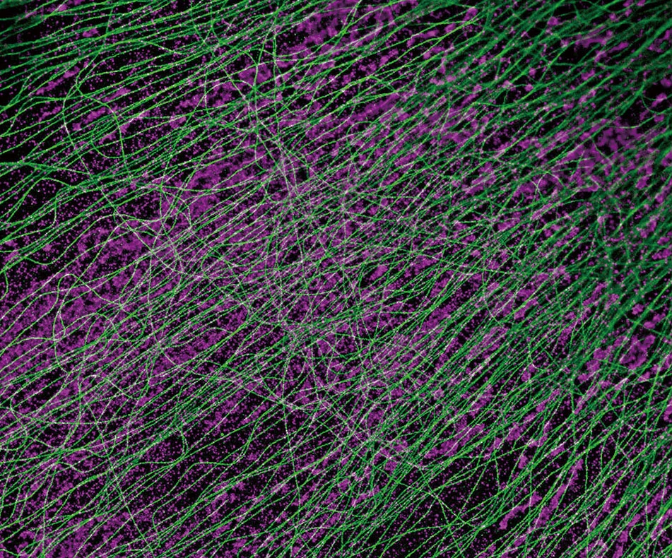
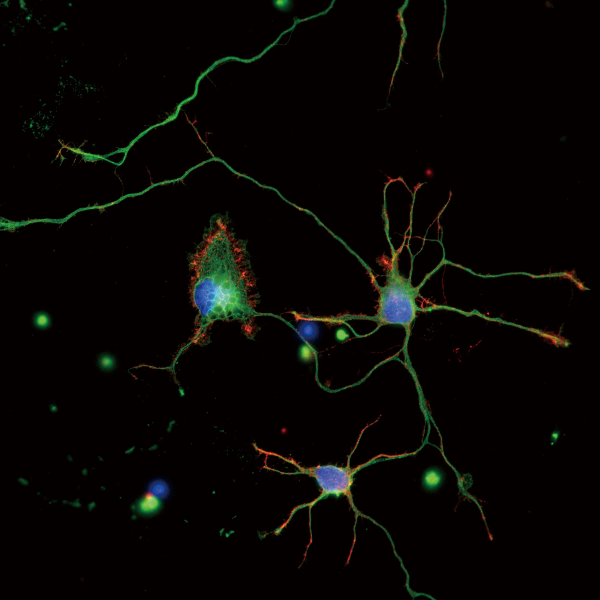
High-Resolution Imaging of Neurons and CV-1 cells expressing α -tubulin (green) and TOMM-20 (magenta) captured with Nikon Microscopy Systems
Nikon microscopes, combined with high-speed cameras and the NIS-Elements imaging software, offer comprehensive control over image acquisition—from basic to advanced levels. The NIS-Elements software integrates powerful analysis tools and impressive visualization capabilities. This combination opens up extensive customization options to support a wide range of your research applications.
Integration of Microscopes with Micromanipulator Systems
CYTOLOGY: FROM ONCOLOGY TO CELL INTERACTIONS
High-resolution confocal images of human malignant osteosarcoma cells, showcasing detailed cellular structures
Nikon microscopes stand out for their flexible adaptability to specialized cytological applications, particularly in cancer research. The system supports precise analysis of nuclear morphology, chromosomal abnormalities, and cellular malignancy features, facilitating early detection of carcinogenesis and micro-level evaluation of treatment efficacy.
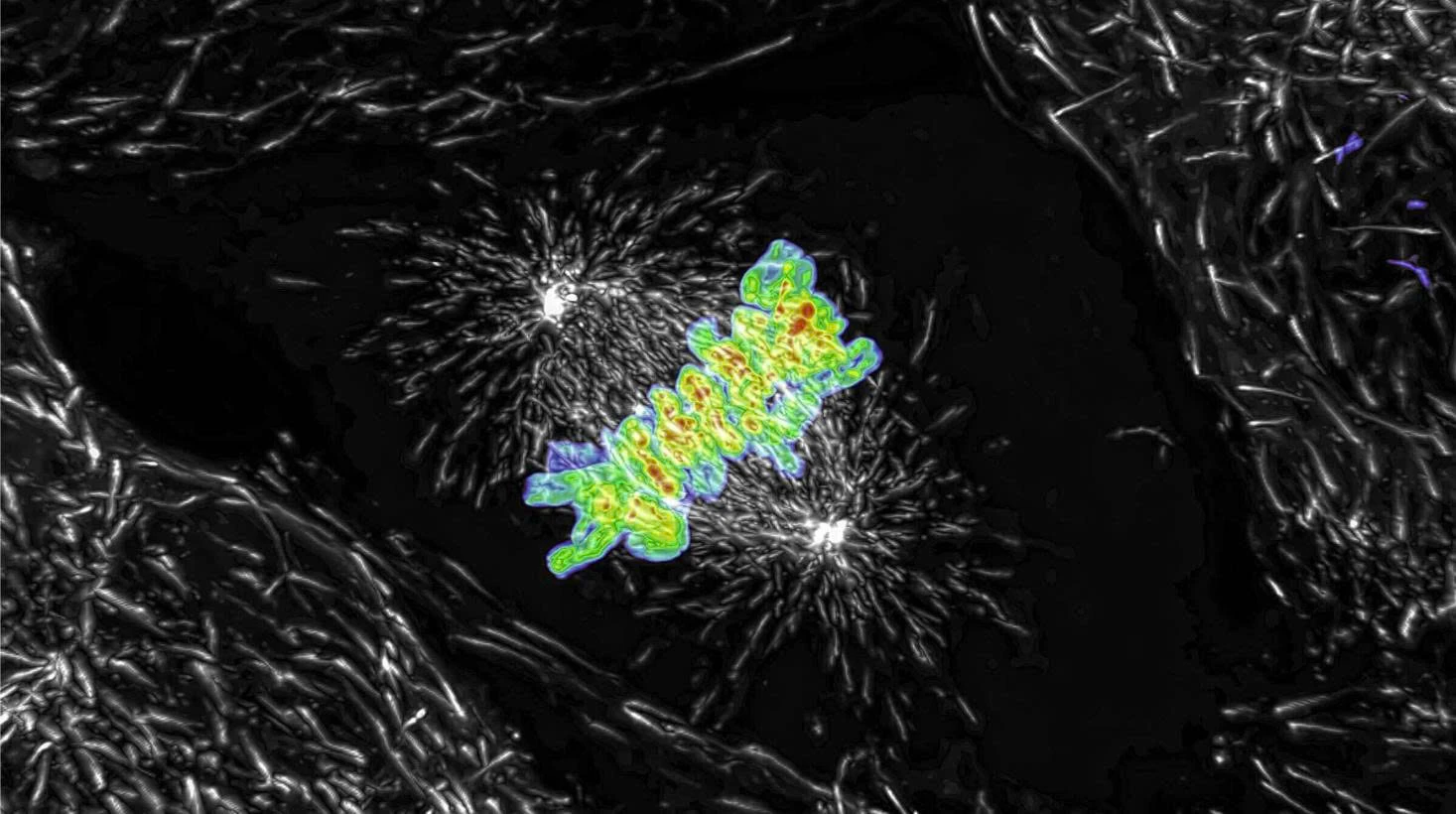
Images of live mitotic cells captured using the AX R system.
Nikon microscopes support tracking of cell division and differentiation processes. Combined with 3D reconstruction technology (Z-stack), they enable spatial modeling of cell cluster development—an essential factor in regenerative medicine research and cell therapy.
Nikon microscopes are favored in virology research for their ability to capture the process of viral entry and its effects on host cells. Notably, the time-lapse imaging mode combined with live/dead staining effectively supports cell toxicity assessment in preclinical studies.
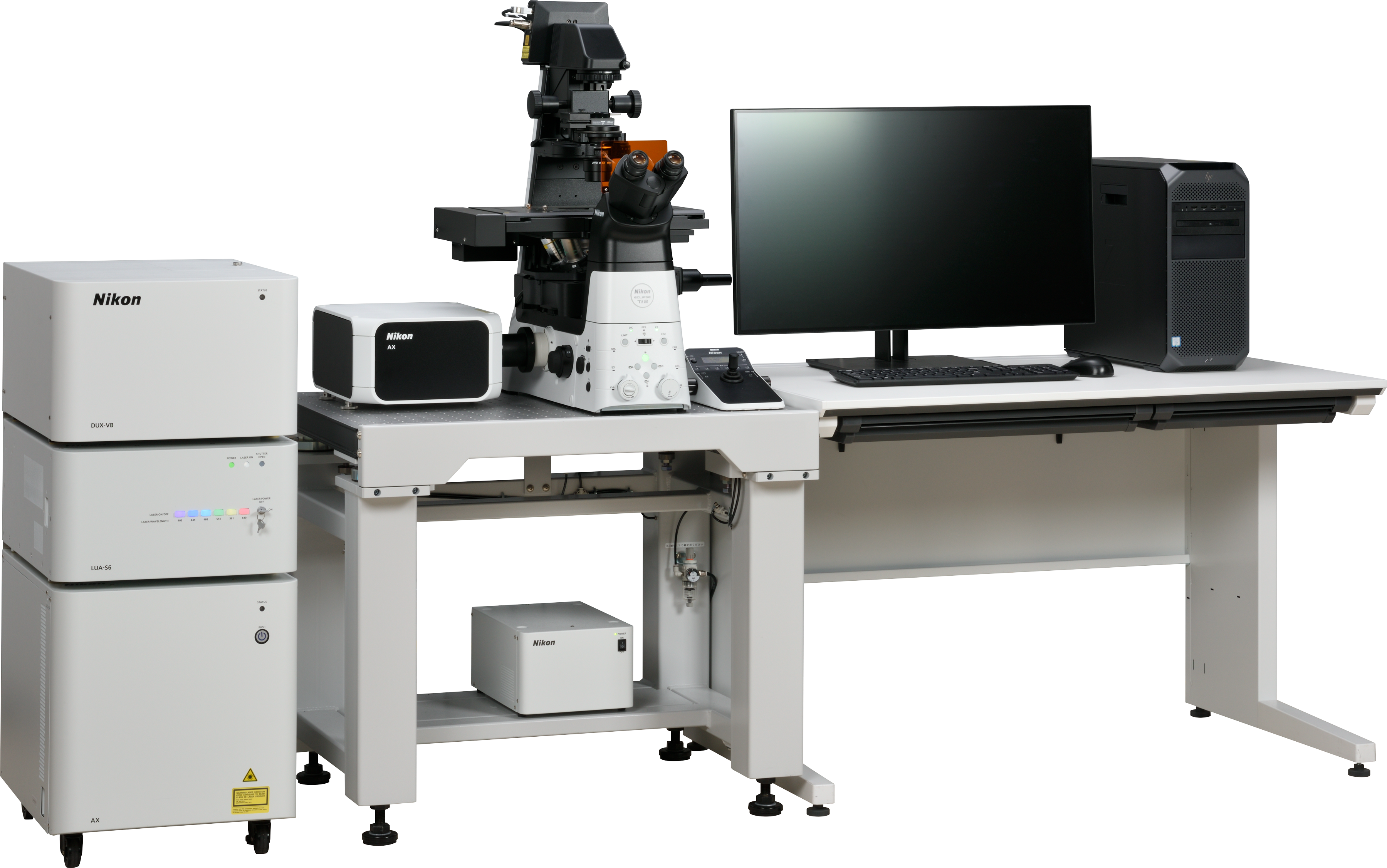
CONFOCAL SYSTEM
As the top choice for modern laboratories, Nikon’s confocal microscopy systems perfectly combine cutting-edge optical technology with powerful image analysis capabilities, setting a new standard in molecular-level life science research.
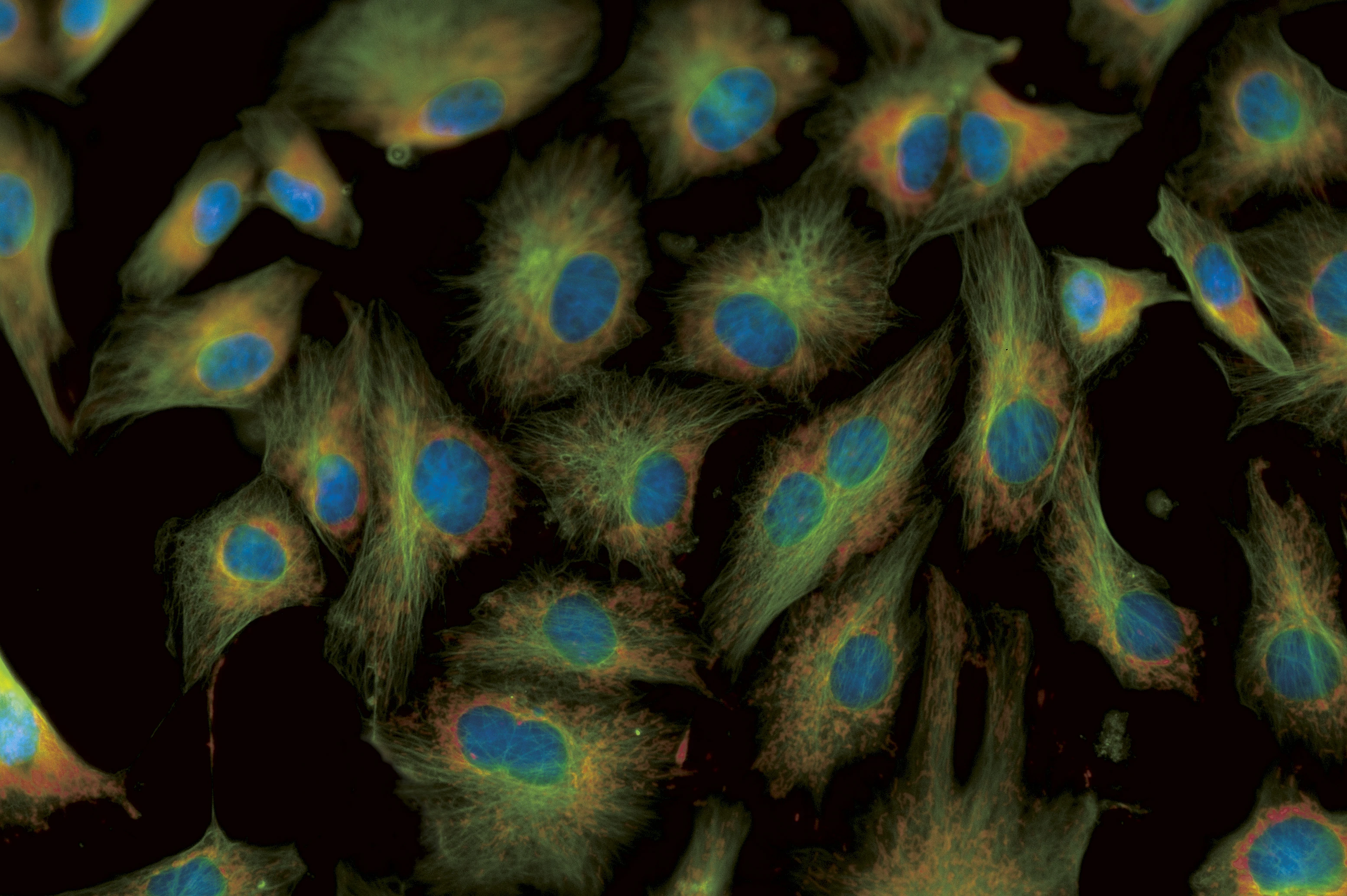
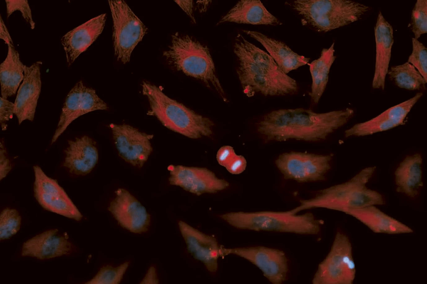
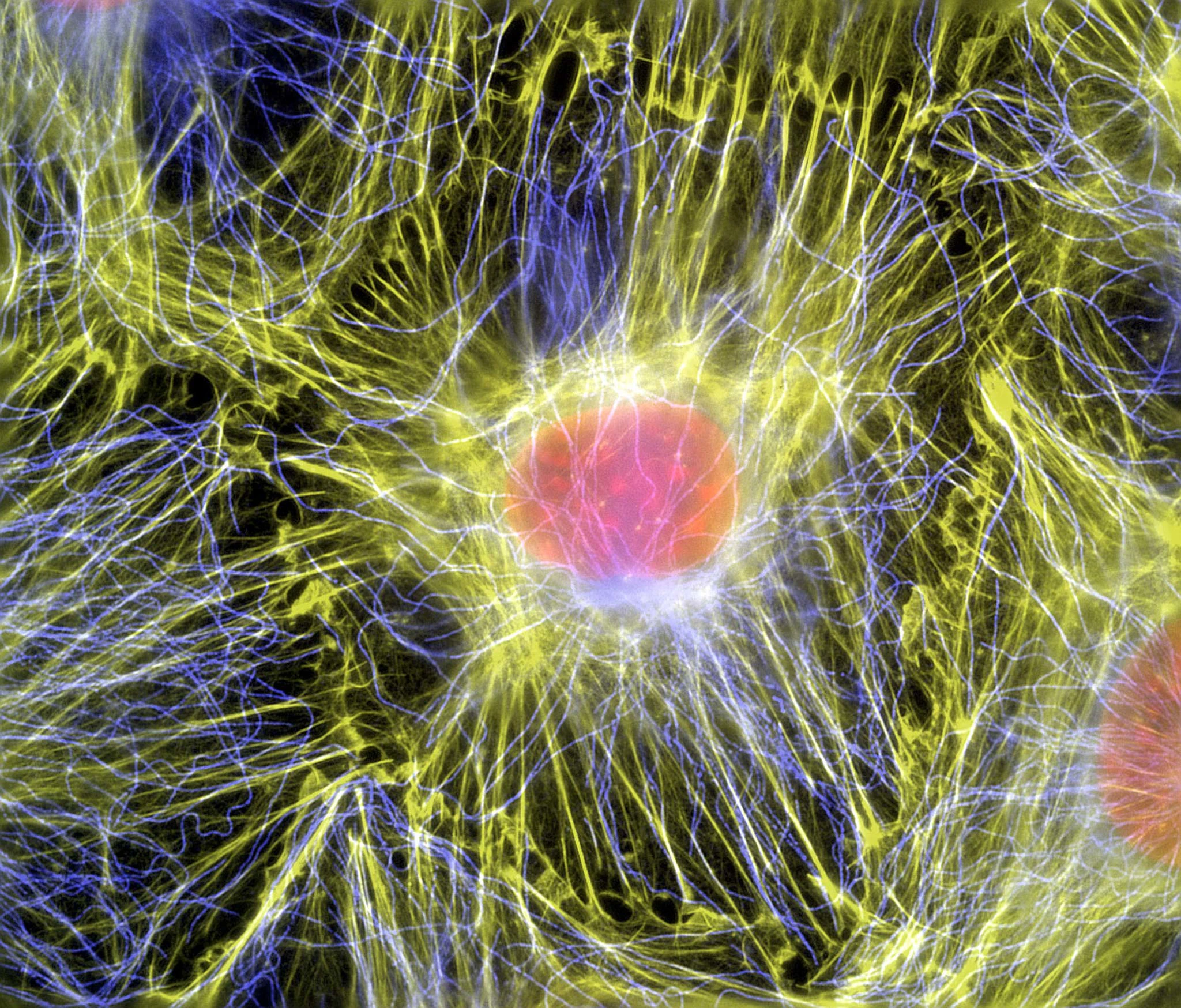
Filamentous actin and microtubules (structural proteins) in mouse fibroblasts (cells)
See at the molecular level with super-resolution imaging enabled by advanced laser scanning technology
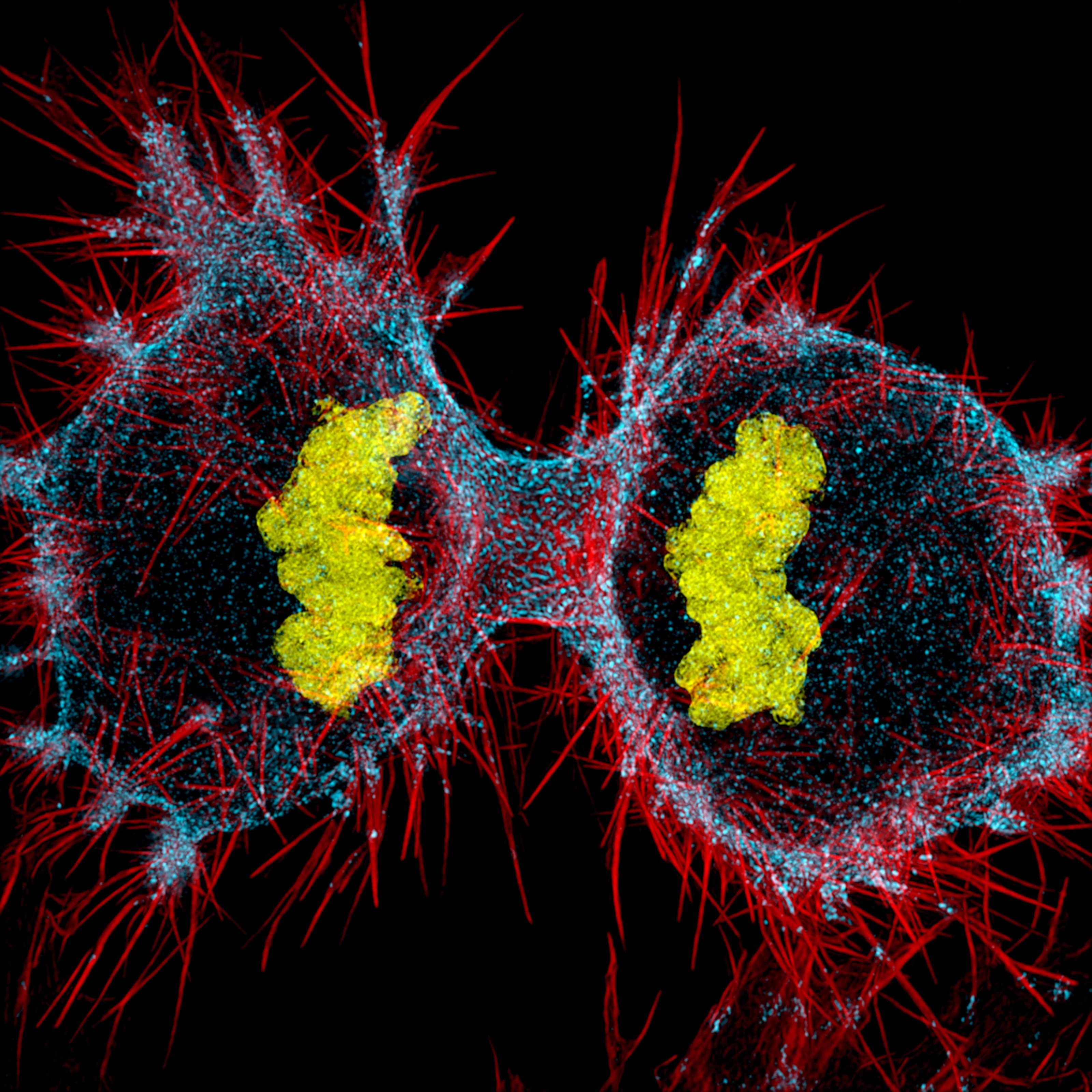
Human HeLa cell undergoing cell division (cytokinesis). DNA (yellow), myosin II (blue) and actin filaments (red)
Observe live specimens and cell interactions with real-time live-cell tracking, enabling accurate and stable recording of biological processes.
Maximize work efficiency with intuitive software featuring powerful functions, supporting 4D/5D image processing, automated measurements, and quantitative analysis.
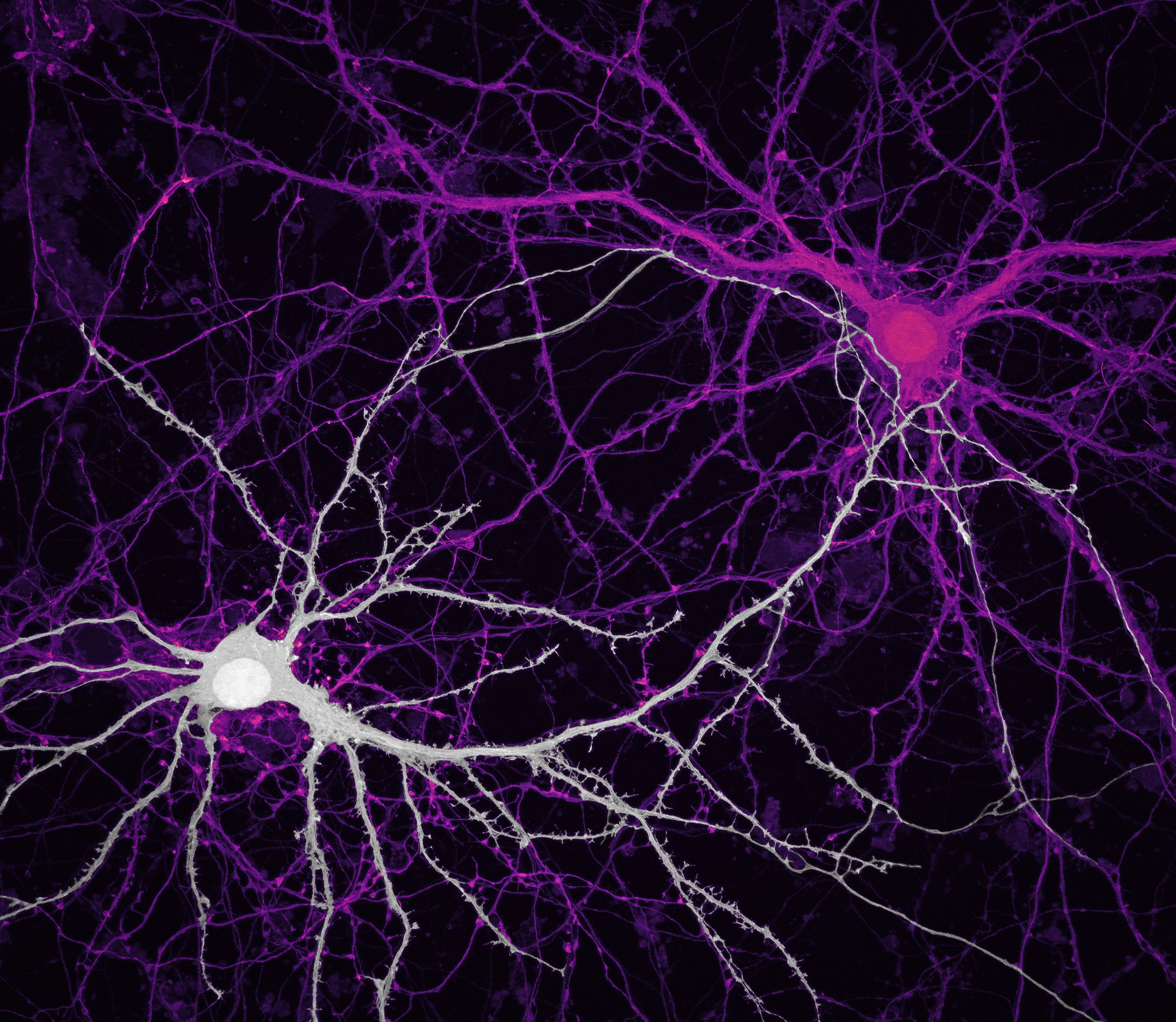
Connections between hippocampal neurons (brain cells)
Nikon confocal systems support simultaneous detection of multiple fluorescence signals with high sensitivity and ultra-low noise, making them ideal for multichannel applications in oncology, immunology, and neuroscience.
Enable limitless research possibilities with versatile upgradeability and customization options.
With over 100 years of expertise in optics, Nikon microscopes deliver not only sharp imaging but also powerful tools for quantitative analysis, molecular mechanism research, and biomedical technology development. In the field of cytology—where every tiny detail can hold groundbreaking value—Nikon stands as the trusted companion for scientists.